Zhang Hongxue, Du Jianwen, Wang Hong, Wang Haili, Jiang Jianhui, Zhao Jingjie, Lu Huan
Department of UItrasonic Diagnosis, The Second Clinic, Institute of the Chengde Medical College, Chengde, Hebei 067000, P.R. China.
Department of Pathology, The Second Clinic, Institute of the Chengde Medical College, Chengde, Hebei 067000, P.R. China.
Exp Ther Med. 2017 Jul;14(1):680-688. doi: 10.3892/etm.2017.4525. Epub 2017 May 31.
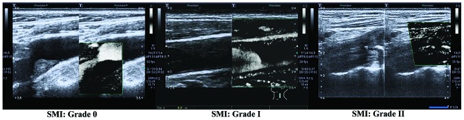
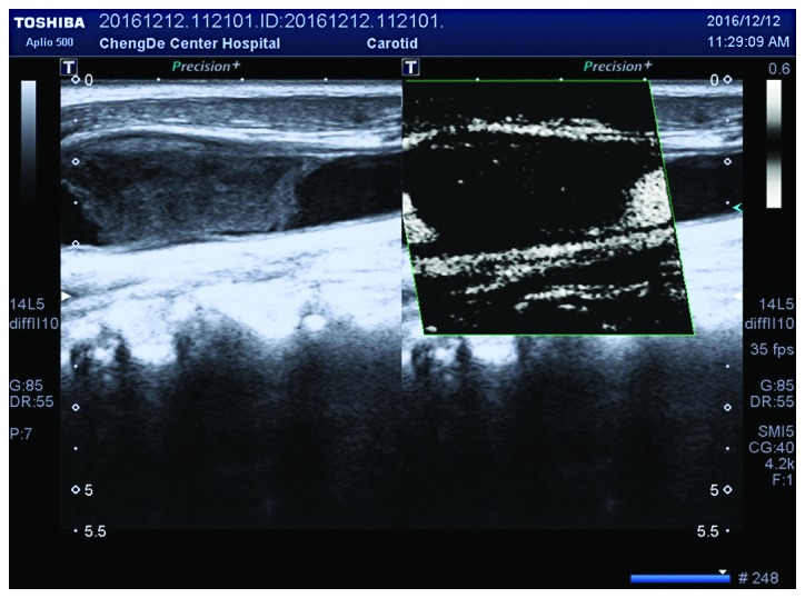
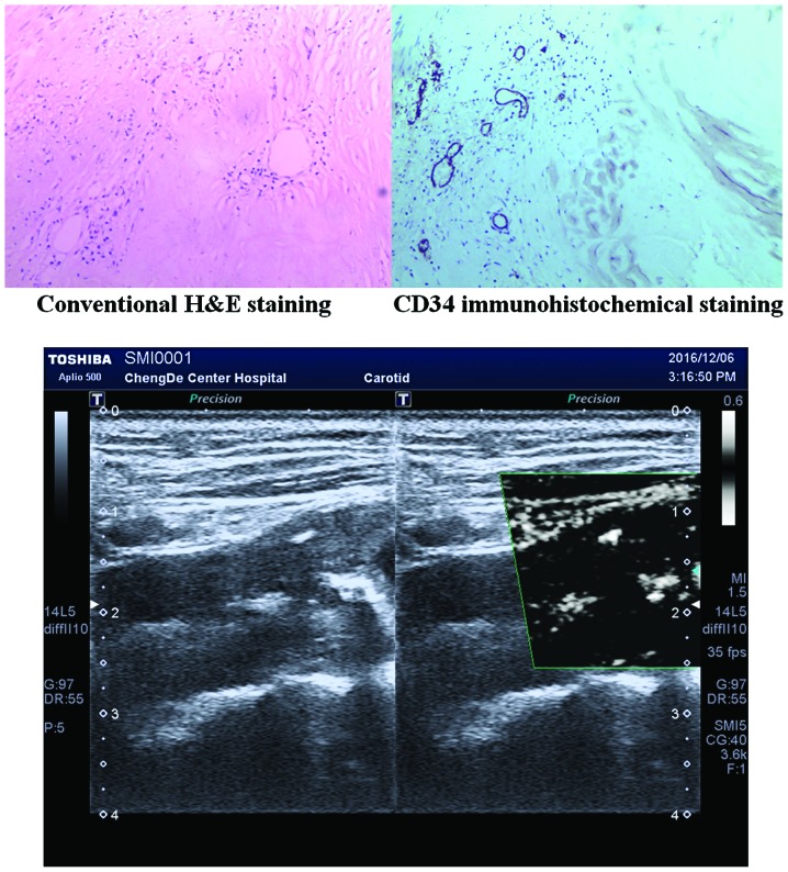
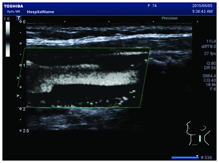
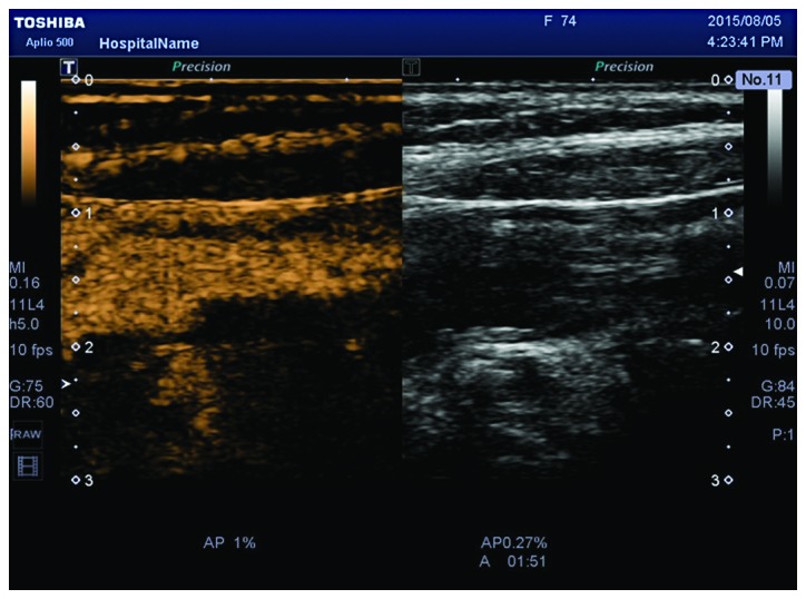
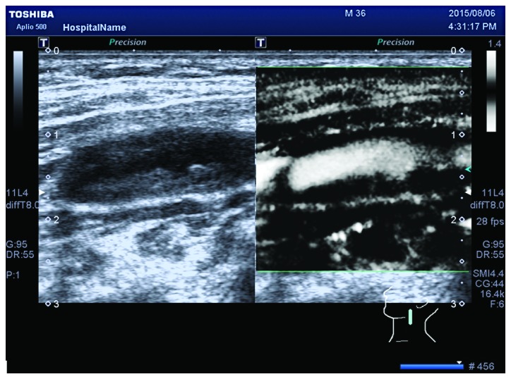
The aim of the present study was to compare the diagnostic values of ultrasound micro-flow imaging (SMI) and contrast-enhanced ultrasound (CEUS) for neovascularization in carotid plaques, and to investigate their capacities for predicting the risks of cerebral stroke. A total of 39 patients (64 carotid plaques) with severe carotid artery stenosis undergoing carotid endarterectomy were selected between February 2015 and February 2016, and SMI and CEUS were used to detect neovascularization in plaques. According to the CEUS dynamic graph of plaques, the enhanced intensity visual scales and contrast parameters were obtained. Carotid atherosclerotic plaques were divided into 4 groups. The differences in the enhanced intensity visual scales, contrast parameters, and gray-scale median (GSM) values among the 4 groups were analyzed. Carotid plaque tissue samples from patients were stained for CD34, and the consistency of the methods for the diagnosis of neovascularization in plaques was analyzed. The differences in GSM values, enhanced intensities, and enhanced densities among the 4 groups of plaques were statistically significant (F=29.365, χ=29.025, χ=30.871, P<0.001); the differences in enhanced intensities of carotid atherosclerotic plaques with different echo types were statistically significant (χ=17.951, P<0.001). The enhanced intensity of plaques was negatively correlated with the GSM value (r=-0.376, P<0.01), and the enhanced density of plaques was negatively correlated with the GSM value (r=-0.252, P<0.01). SMI and CEUS grading had good consistency (κ=0.860>0), there were statistically significant differences in new vessel densities with different SMI gradings (P<0.001), and the clinical symptoms and severity were positively correlated with SMI grading (rs=0.592>0). In conclusion, SMI and CEUS have good consistency for evaluating neovascularization in carotid plaques, and have good clinical value for evaluating neovascularization in carotid plaques.
本研究旨在比较超声微血管成像(SMI)和超声造影(CEUS)对颈动脉斑块新生血管形成的诊断价值,并探讨它们预测脑卒风险的能力。2015年2月至2016年2月期间,共选取了39例接受颈动脉内膜切除术的重度颈动脉狭窄患者(64个颈动脉斑块),采用SMI和CEUS检测斑块内新生血管形成。根据斑块的CEUS动态图,获取增强强度视觉评分和造影参数。将颈动脉粥样硬化斑块分为4组。分析4组之间增强强度视觉评分、造影参数和灰度中位数(GSM)值的差异。对患者的颈动脉斑块组织样本进行CD34染色,分析斑块内新生血管形成诊断方法的一致性。4组斑块的GSM值、增强强度和增强密度差异有统计学意义(F=29.365,χ=29.025,χ=30.871,P<0.001);不同回声类型的颈动脉粥样硬化斑块增强强度差异有统计学意义(χ=17.951,P<0.001)。斑块的增强强度与GSM值呈负相关(r=-0.376,P<0.01),斑块的增强密度与GSM值呈负相关(r=-0.252,P<0.01)。SMI和CEUS分级具有良好的一致性(κ=0.860>0),不同SMI分级的新生血管密度差异有统计学意义(P<0.001),临床症状和严重程度与SMI分级呈正相关(rs=0.592>0)。总之,SMI和CEUS在评估颈动脉斑块新生血管形成方面具有良好的一致性,对评估颈动脉斑块新生血管形成具有良好的临床价值。