Rosa A R, Steffens D, Santi B, Quintiliano K, Steffen N, Pilger D A, Pranke P
Laboratório de Hematologia e Células Tronco, Faculdade de Farmácia, Universidade Federal do Rio Grande do Sul, Porto Alegre, RS, Brasil.
Programa de Pós-graduação em Ciência dos Materiais, Universidade Federal do Rio Grande do Sul, Porto Alegre, RS, Brasil.
Braz J Med Biol Res. 2017 Aug 7;50(9):e5648. doi: 10.1590/1414-431X20175648.
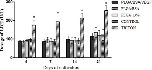
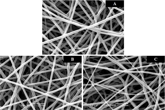
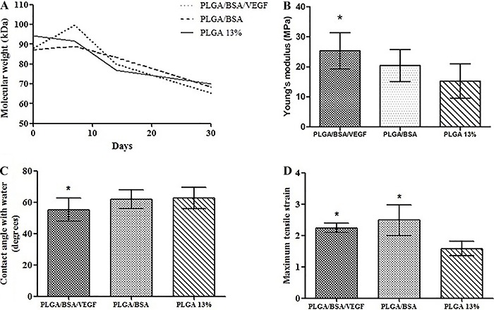
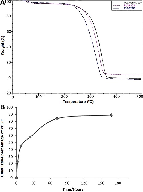
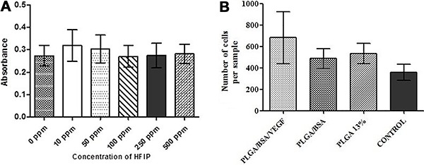
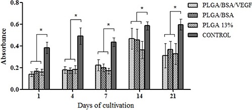
The association of bioactive molecules, such as vascular endothelial growth factor (VEGF), with nanofibers facilitates their controlled release, which could contribute to cellular migration and differentiation in tissue regeneration. In this research, the influence of their incorporation on a polylactic-co-glycolic acid (PLGA) scaffold produced by electrospinning on cell adhesion and viability and cytotoxicity was carried out in three groups: 1) PLGA/BSA/VEGF; 2) PLGA/BSA, and 3) PLGA. Morphology, fiber diameter, contact angle, loading efficiency and controlled release of VEGF of the biomaterials, among others, were measured. The nanofibers showed smooth surfaces without beads and with interconnected pores. PLGA/BSA/VEGF showed the smallest water contact angle and VEGF released for up to 160 h. An improvement in cell adhesion was observed for the PLGA/BSA/VEGF scaffolds compared to the other groups and the scaffolds were non-toxic for the cells. Therefore, the scaffolds were shown to be a good strategy for sustained delivery of VEGF and may be a useful tool for tissue engineering.
生物活性分子,如血管内皮生长因子(VEGF),与纳米纤维的结合有助于其控释,这可能有助于组织再生中的细胞迁移和分化。在本研究中,通过静电纺丝制备的聚乳酸-乙醇酸共聚物(PLGA)支架中加入这些分子对细胞黏附、活力和细胞毒性的影响在三组中进行:1)PLGA/牛血清白蛋白/VEGF;2)PLGA/牛血清白蛋白;3)PLGA。测量了生物材料的形态、纤维直径、接触角、VEGF的负载效率和控释等。纳米纤维表面光滑无珠且有相互连通的孔隙。PLGA/牛血清白蛋白/VEGF的水接触角最小,VEGF释放长达160小时。与其他组相比,PLGA/牛血清白蛋白/VEGF支架的细胞黏附有所改善,且该支架对细胞无毒。因此,这些支架被证明是VEGF持续递送的良好策略,可能是组织工程的有用工具。