Lecka-Czernik Beata, Stechschulte Lance A, Czernik Piotr J, Sherman Shermel B, Huang Shilong, Krings Amrei
Department of Orthopaedic Surgery, University of Toledo Health Sciences Campus, Toledo, OH, United States.
Department of Physiology and Pharmacology, University of Toledo Health Sciences Campus, Toledo, OH, United States.
Front Endocrinol (Lausanne). 2017 Aug 4;8:188. doi: 10.3389/fendo.2017.00188. eCollection 2017.
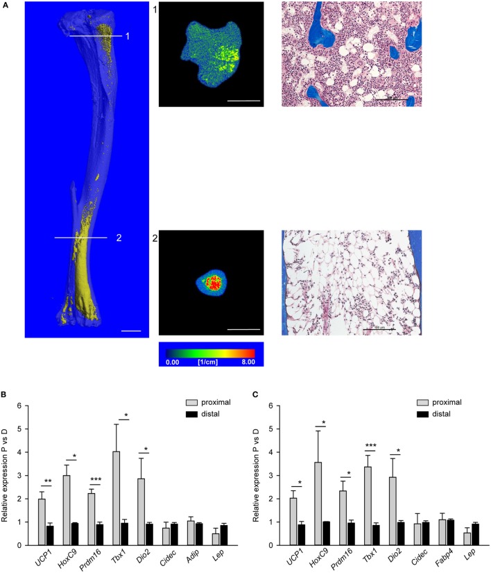
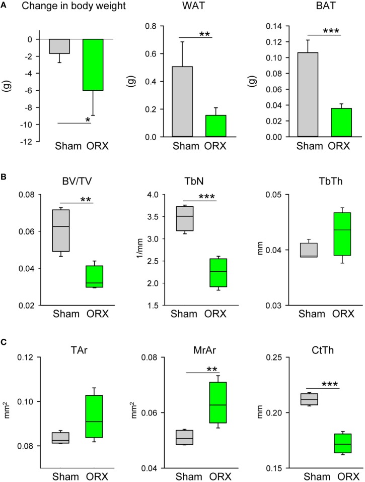
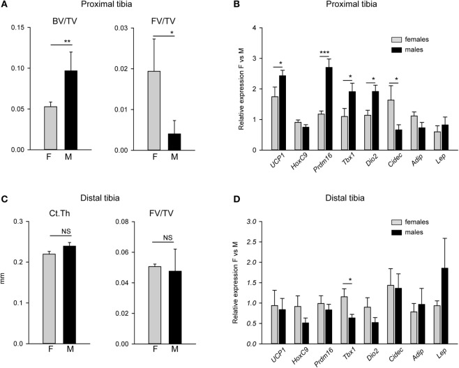
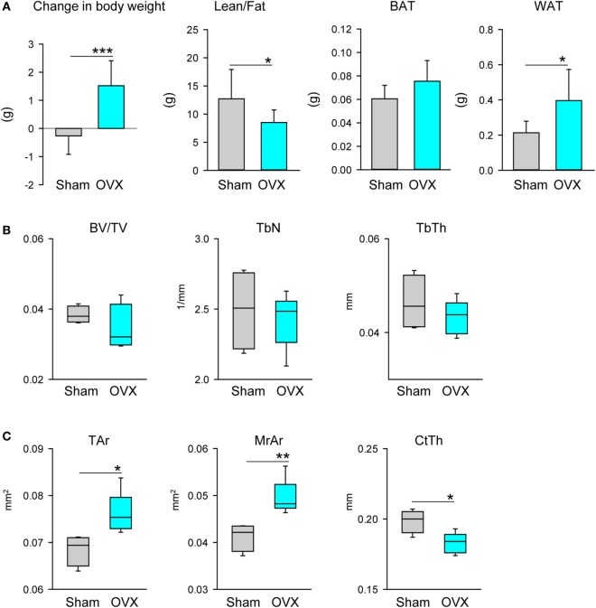
Marrow adipose tissue (MAT) is unique with respect to origin, metabolism, and function. MAT is characterized with high heterogeneity which correlates with skeletal location and bone metabolism. This fat depot is also highly sensitive to various hormonal, environmental, and pharmacologic cues to which it responds with changes in volume and/or metabolic phenotype. We have demonstrated previously that MAT has characteristics of both white (WAT) and brown (BAT)-like or beige adipose tissue, and that beige phenotype is attenuated with aging and in diabetes. Here, we extended our analysis by comparing MAT phenotype in different locations within a tibia bone of mature C57BL/6 mice and with respect to the presence of sex steroids in males and females. We report that MAT juxtaposed to trabecular bone of proximal tibia (pMAT) is characterized by elevated expression of beige fat markers including , and , when compared with MAT located in distal tibia (dMAT). There is also a difference in tissue organization with adipocytes in proximal tibia being dispersed between trabeculae, while adipocytes in distal tibia being densely packed. Higher trabecular bone mass (BV/TV) in males correlates with lower pMAT volume and higher expression of beige markers in the same location, when compared with females. However, there is no sexual divergence in the volume and transcriptional profile of dMAT. A removal of ovaries in females resulted in decreased cortical bone mass and increased volume of both pMAT and dMAT, as well as volume of gonadal WAT (gWAT). Increase in pMAT volume was associated with marked increase in and expression and relative decrease in beige fat gene markers. A removal of testes in males resulted in cortical and trabecular bone loss and the tendency to increased volume of both pMAT and dMAT, despite a loss of gWAT. Orchiectomy did not affect the expression of white and beige adipocyte gene markers. In conclusion, expression profile of beige adipocyte gene markers correlates with skeletal location of active bone remodeling and higher BV/TV, however bone loss resulted from sex steroid deficiency is not proportional to MAT expansion at the same skeletal location.
骨髓脂肪组织(MAT)在起源、代谢和功能方面具有独特性。MAT具有高度异质性,这与骨骼位置和骨代谢相关。这个脂肪库对各种激素、环境和药理信号也高度敏感,并会通过体积和/或代谢表型的变化做出反应。我们之前已经证明,MAT具有白色脂肪组织(WAT)和棕色脂肪组织(BAT)样或米色脂肪组织的特征,并且米色表型会随着衰老和糖尿病而减弱。在这里,我们通过比较成熟C57BL/6小鼠胫骨不同部位的MAT表型以及雄性和雌性中是否存在性类固醇,扩展了我们的分析。我们报告称,与位于胫骨远端(dMAT)的MAT相比,与胫骨近端小梁骨并列的MAT(pMAT)的特征是米色脂肪标记物(包括 、 和 )的表达升高。组织结构也存在差异,胫骨近端的脂肪细胞分散在小梁之间,而胫骨远端的脂肪细胞则紧密堆积。与雌性相比,雄性中较高的小梁骨量(BV/TV)与相同位置较低的pMAT体积和较高的米色标记物表达相关。然而,dMAT的体积和转录谱没有性别差异。雌性去卵巢导致皮质骨量减少,pMAT和dMAT的体积以及性腺白色脂肪组织(gWAT)的体积增加。pMAT体积的增加与 和 表达的显著增加以及米色脂肪基因标记物的相对减少相关。雄性去睾丸导致皮质骨和小梁骨丢失,尽管gWAT减少,但pMAT和dMAT的体积有增加的趋势。睾丸切除术不影响白色和米色脂肪细胞基因标记物的表达。总之,米色脂肪细胞基因标记物的表达谱与活跃骨重塑的骨骼位置和较高的BV/TV相关,然而,性类固醇缺乏导致的骨质流失与同一骨骼位置的MAT扩张不成比例。