Zhou Heling, Hallac Rami R, Yuan Qing, Ding Yao, Zhang Zhongwei, Xie Xian-Jin, Francis Franto, Roehrborn Claus G, Sims R Douglas, Costa Daniel N, Raj Ganesh V, Mason Ralph P
Department of Radiology, University of Texas Southwestern Medical Center, Dallas, TX 75390, USA.
Analytical Imaging and Modeling Center, Children's Medical Center, Dallas, TX 75235, USA.
Diagnostics (Basel). 2017 Aug 24;7(3):48. doi: 10.3390/diagnostics7030048.
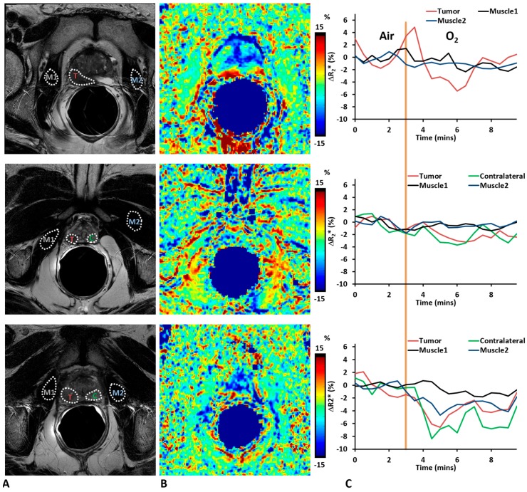
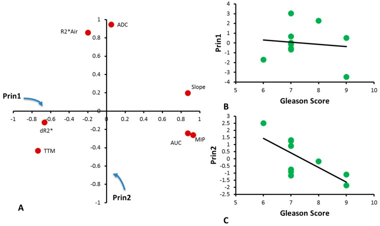
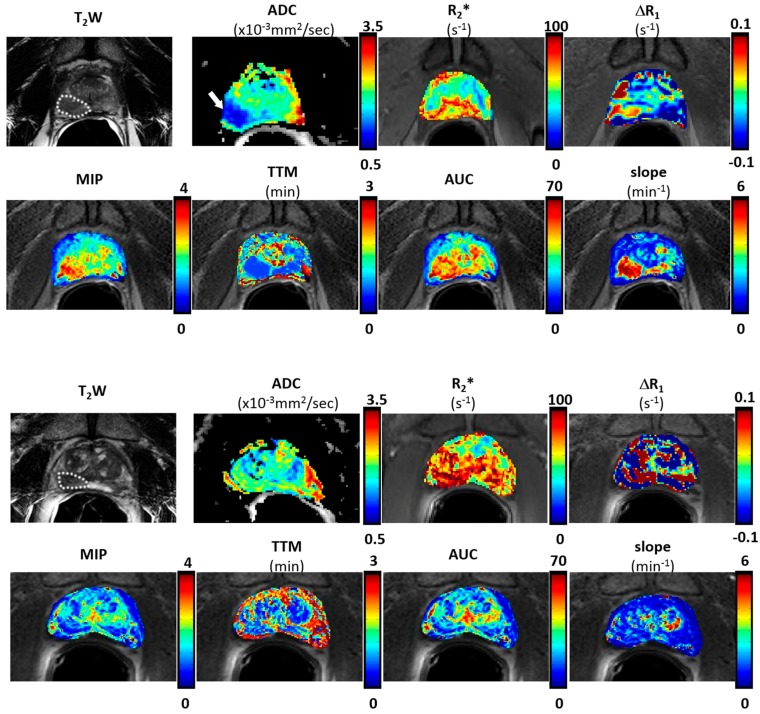
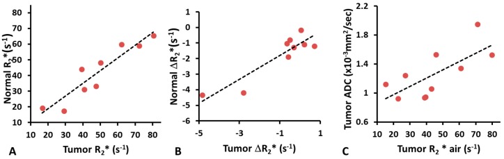
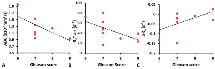
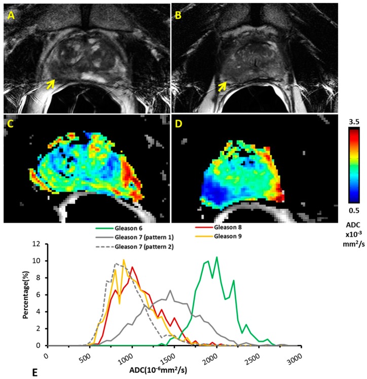
Hypoxia is associated with prostate tumor aggressiveness, local recurrence, and biochemical failure. Magnetic resonance imaging (MRI) offers insight into tumor pathophysiology and recent reports have related transverse relaxation rate (R₂*) and longitudinal relaxation rate (R₁) measurements to tumor hypoxia. We have investigated the inclusion of oxygen-enhanced MRI for multi-parametric evaluation of tumor malignancy. Multi-parametric MRI sequences at 3 Tesla were evaluated in 10 patients to investigate hypoxia in prostate cancer prior to radical prostatectomy. Blood oxygen level dependent (BOLD), tissue oxygen level dependent (TOLD), dynamic contrast enhanced (DCE), and diffusion weighted imaging MRI were intercorrelated and compared with the Gleason score. The apparent diffusion coefficient (ADC) was significantly lower in tumor than normal prostate. Baseline R₂* (BOLD-contrast) was significantly higher in tumor than normal prostate. Upon the oxygen breathing challenge, R₂* decreased significantly in the tumor tissue, suggesting improved vascular oxygenation, however changes in R₁ were minimal. R₂* of contralateral normal prostate decreased in most cases upon oxygen challenge, although the differences were not significant. Moderate correlation was found between ADC and Gleason score. ADC and R₂* were correlated and trends were found between Gleason score and R₂*, as well as maximum-intensity-projection and area-under-the-curve calculated from DCE. Tumor ADC and R₂* have been associated with tumor hypoxia, and thus the correlations are of particular interest. A multi-parametric approach including oxygen-enhanced MRI is feasible and promises further insights into the pathophysiological information of tumor microenvironment.
缺氧与前列腺肿瘤的侵袭性、局部复发及生化失败相关。磁共振成像(MRI)有助于深入了解肿瘤病理生理学,近期报告已将横向弛豫率(R₂*)和纵向弛豫率(R₁)测量与肿瘤缺氧联系起来。我们研究了将氧增强MRI纳入肿瘤恶性多参数评估的情况。对10例患者进行了3特斯拉的多参数MRI序列评估,以在根治性前列腺切除术前行前列腺癌缺氧情况的研究。血氧水平依赖(BOLD)、组织氧水平依赖(TOLD)、动态对比增强(DCE)及扩散加权成像MRI相互关联,并与Gleason评分进行比较。肿瘤的表观扩散系数(ADC)显著低于正常前列腺。肿瘤的基线R₂*(BOLD对比)显著高于正常前列腺。在吸氧激发试验中,肿瘤组织中的R₂显著降低,提示血管氧合改善,但R₁变化极小。在吸氧激发试验中,大多数情况下对侧正常前列腺的R₂降低,尽管差异不显著。ADC与Gleason评分之间存在中度相关性。ADC与R₂相关,且在Gleason评分与R₂之间以及根据DCE计算的最大强度投影和曲线下面积之间发现了趋势。肿瘤ADC和R₂*与肿瘤缺氧相关,因此这些相关性特别令人关注。包括氧增强MRI的多参数方法是可行的,并有望进一步深入了解肿瘤微环境的病理生理信息。