• 文献检索
  • 文档翻译
  • 深度研究
  • 学术资讯
  • Suppr Zotero 插件Zotero 插件
  • 邀请有礼
  • 套餐&价格
  • 历史记录
应用&插件
Suppr Zotero 插件Zotero 插件浏览器插件Mac 客户端Windows 客户端微信小程序
定价
高级版会员购买积分包购买API积分包
服务
文献检索文档翻译深度研究API 文档MCP 服务
关于我们
关于 Suppr公司介绍联系我们用户协议隐私条款
关注我们

Suppr 超能文献

核心技术专利:CN118964589B侵权必究
粤ICP备2023148730 号-1Suppr @ 2026

文献检索

告别复杂PubMed语法,用中文像聊天一样搜索,搜遍4000万医学文献。AI智能推荐,让科研检索更轻松。

立即免费搜索

文件翻译

保留排版,准确专业,支持PDF/Word/PPT等文件格式,支持 12+语言互译。

免费翻译文档

深度研究

AI帮你快速写综述,25分钟生成高质量综述,智能提取关键信息,辅助科研写作。

立即免费体验

微眼跳采样移动图像信息提供超锐视觉。

Microsaccadic sampling of moving image information provides hyperacute vision.

机构信息

National Key Laboratory of Cognitive Neuroscience and Learning, Beijing Normal University, Beijing, China.

Department of Biomedical Science, University of Sheffield, Sheffield, United Kingdom.

出版信息

Elife. 2017 Sep 5;6:e26117. doi: 10.7554/eLife.26117.

DOI:10.7554/eLife.26117
PMID:28870284
原文链接:https://pmc.ncbi.nlm.nih.gov/articles/PMC5584993/
Abstract

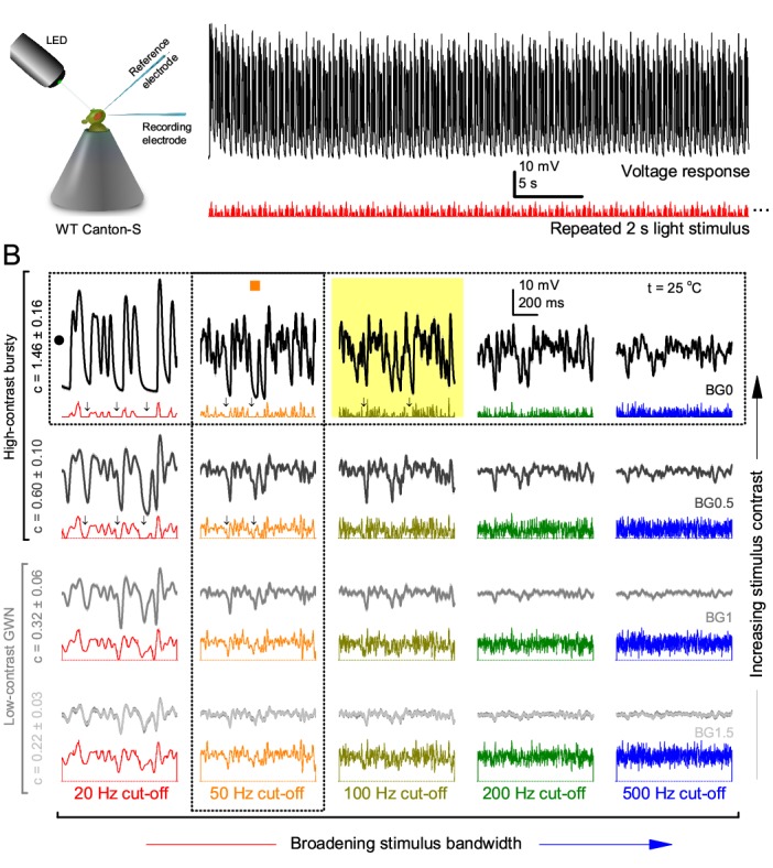
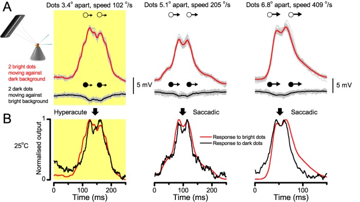
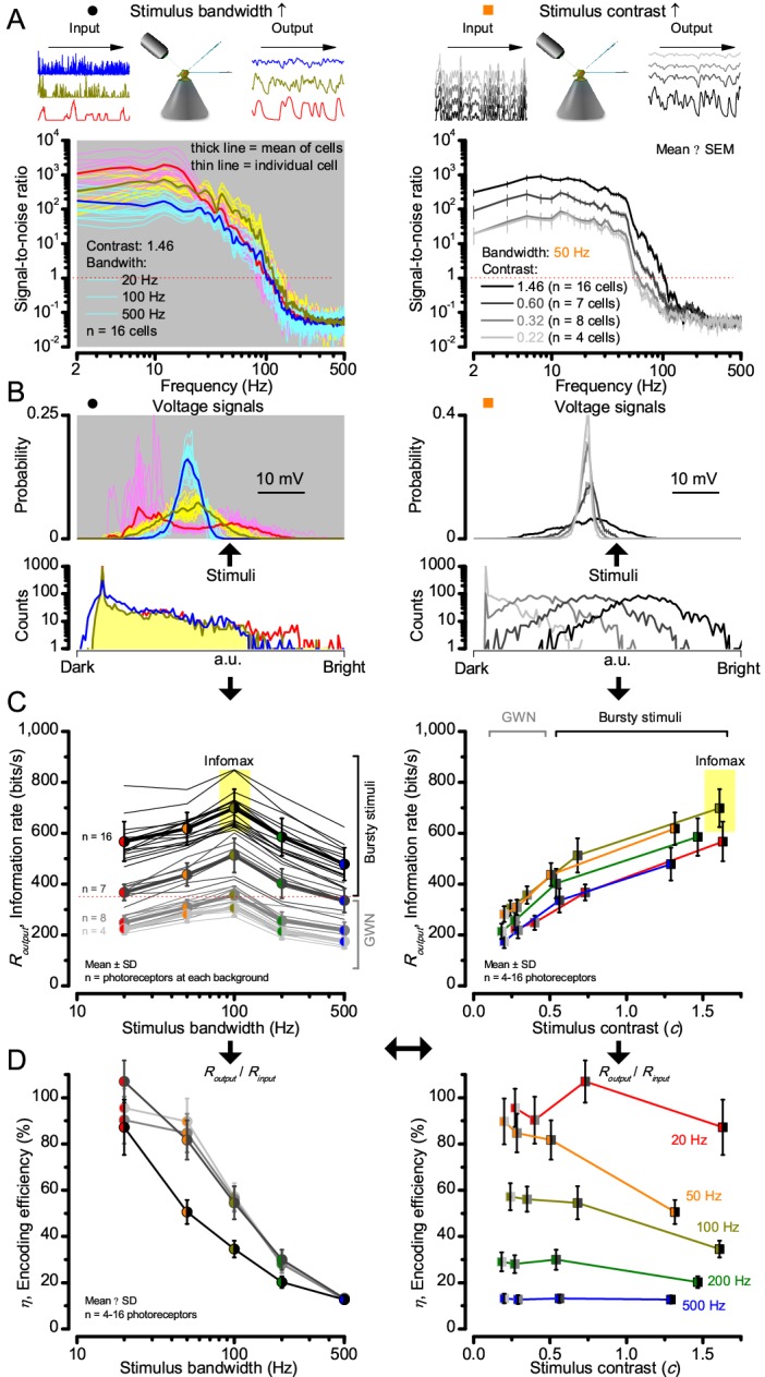
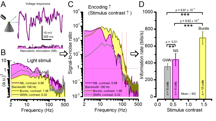
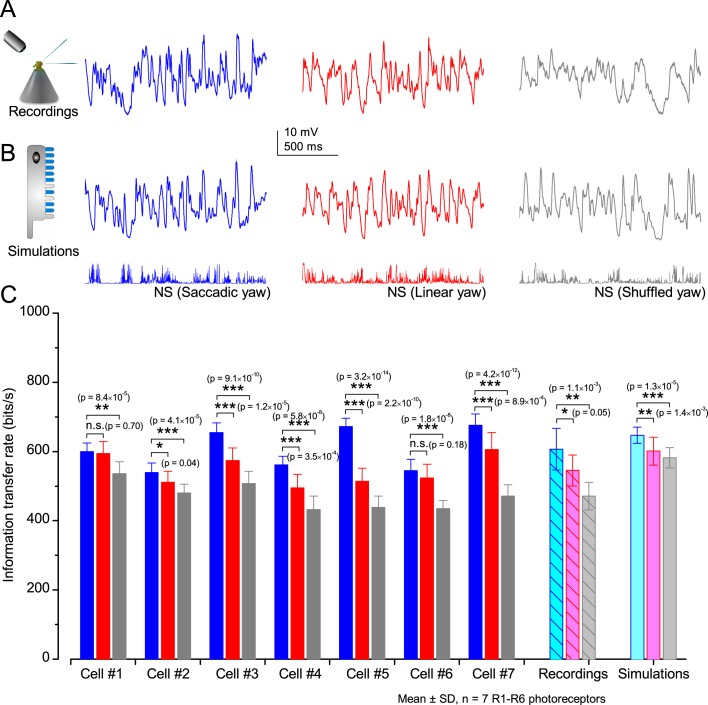
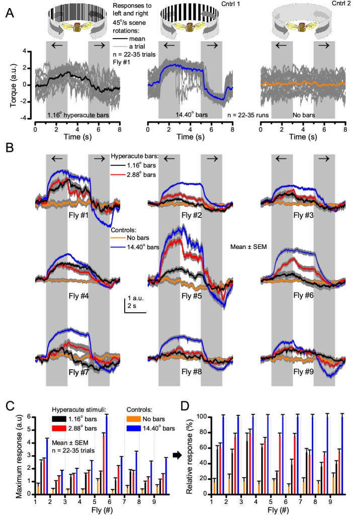
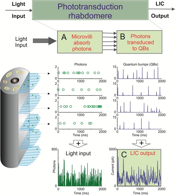
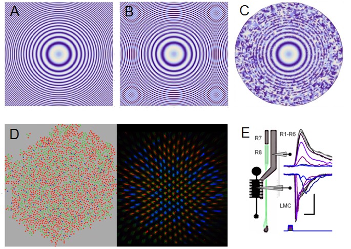
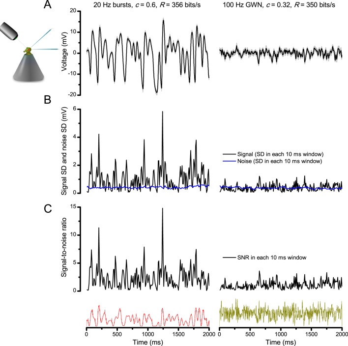
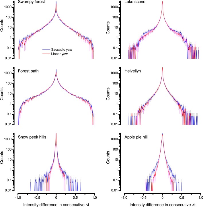
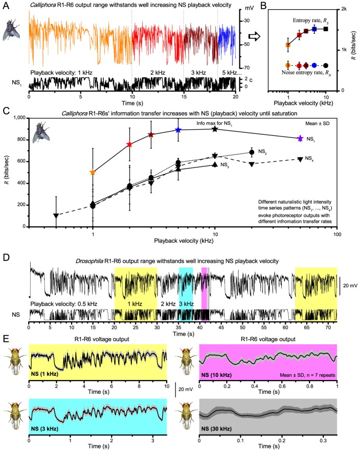
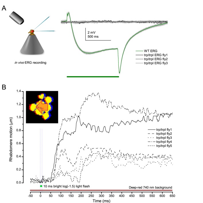
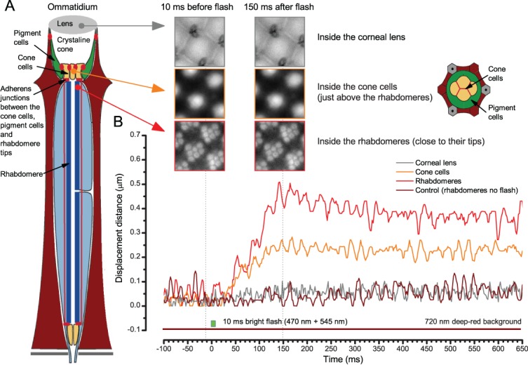
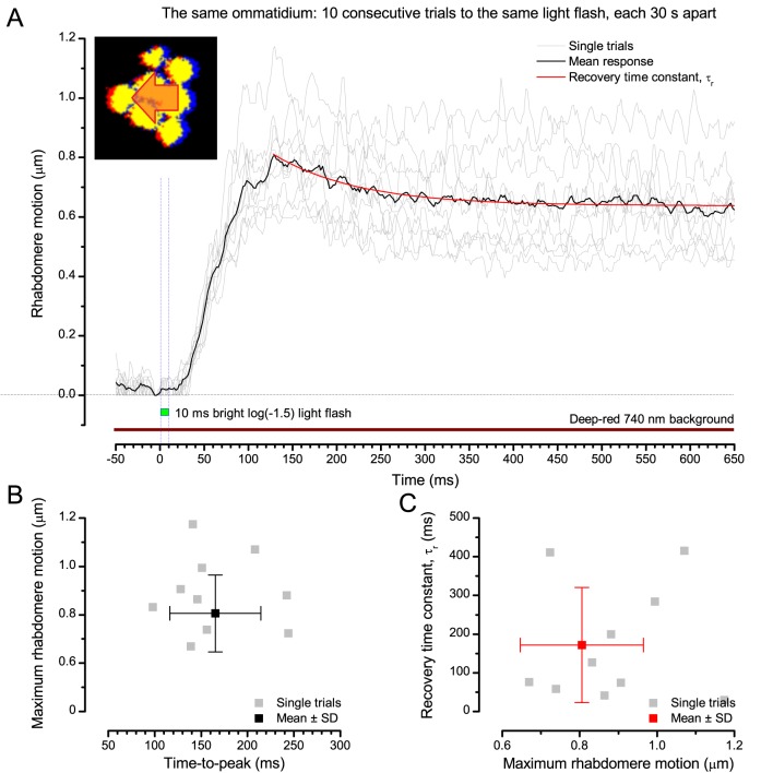
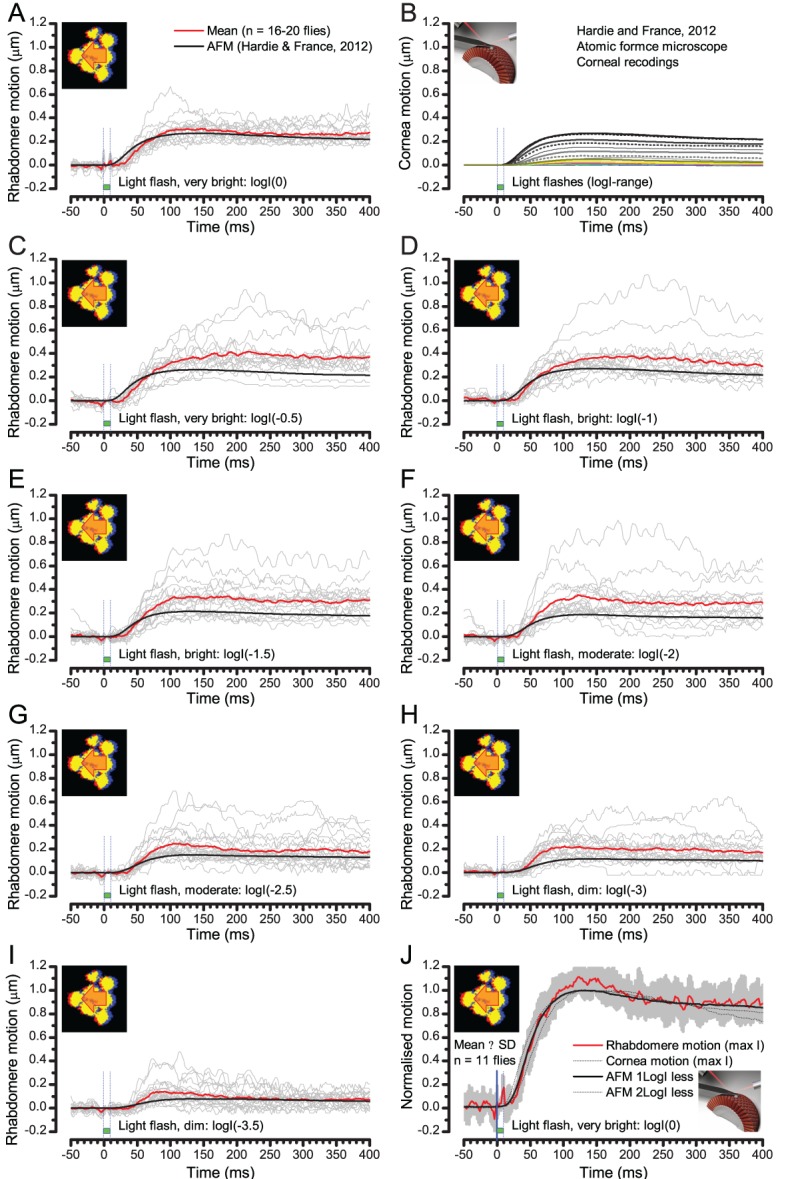
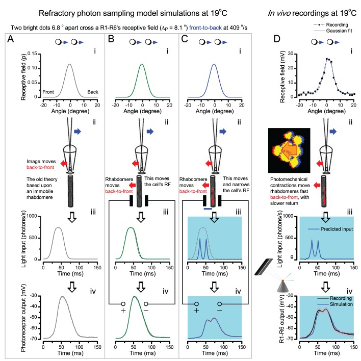
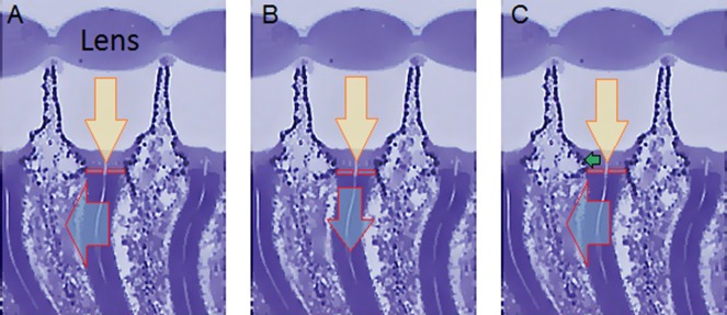
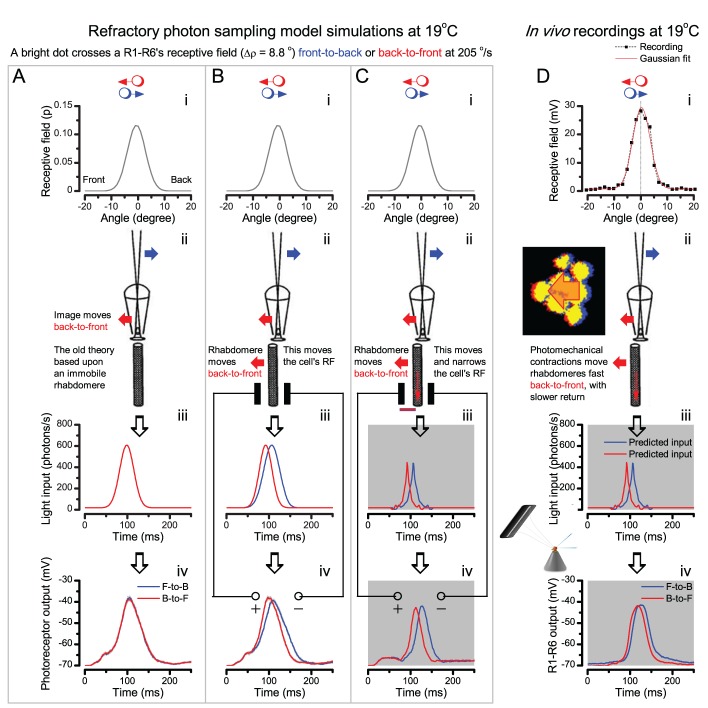
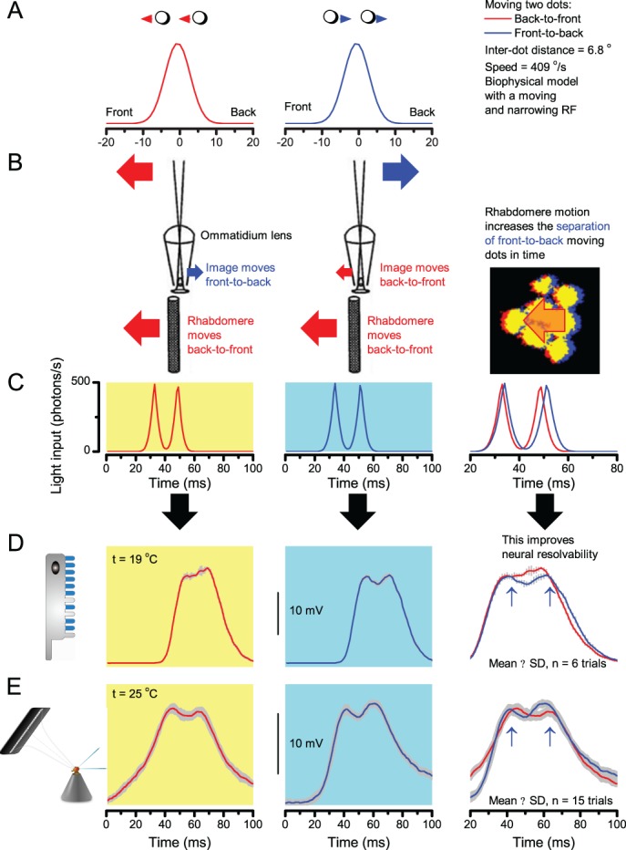
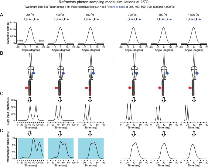
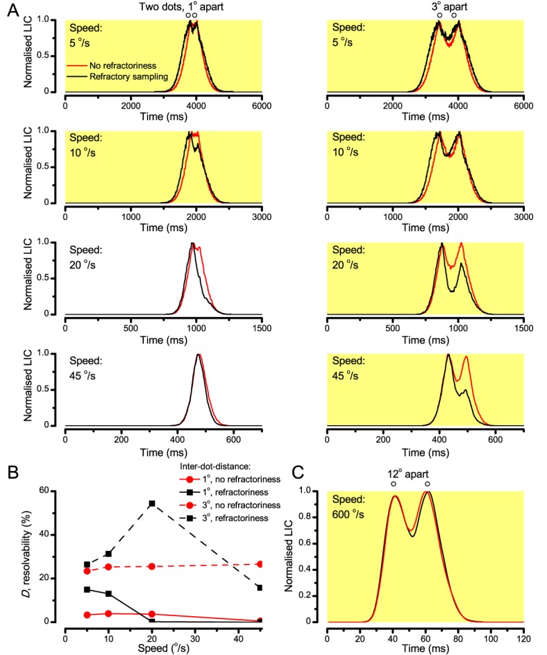
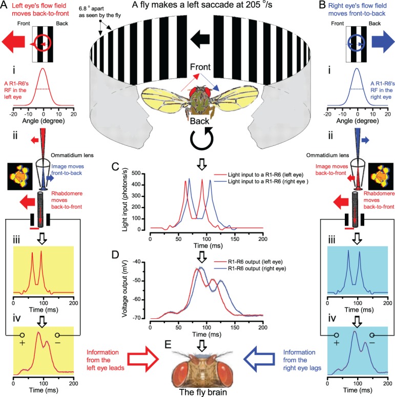
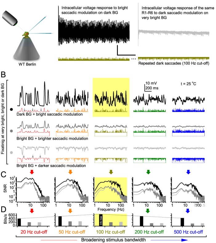
Small fly eyes should not see fine image details. Because flies exhibit saccadic visual behaviors and their compound eyes have relatively few ommatidia (sampling points), their photoreceptors would be expected to generate blurry and coarse retinal images of the world. Here we demonstrate that see the world far better than predicted from the classic theories. By using electrophysiological, optical and behavioral assays, we found that R1-R6 photoreceptors' encoding capacity is maximized to fast high-contrast bursts, which resemble their light input during saccadic behaviors. Whilst , R1-R6s resolve moving objects at saccadic speeds beyond the predicted motion-blur-limit. Our results show how refractory phototransduction and rapid photomechanical photoreceptor contractions jointly sharpen retinal images of moving objects , enabling hyperacute vision, and explain how such microsaccadic information sampling exceeds the compound eyes' optical limits. These discoveries elucidate how acuity depends upon photoreceptor function and eye movements.

摘要

小蝇眼不应看到精细的图像细节。因为蝇类表现出扫视视觉行为,并且它们的复眼相对较少小眼(采样点),它们的光感受器应该会产生模糊和粗糙的世界视网膜图像。在这里,我们证明了它们看到的世界远比经典理论预测的要好。通过使用电生理、光学和行为测定法,我们发现 R1-R6 光感受器的编码能力最大限度地适应于快速高对比度的爆发,这类似于它们在扫视行为期间的光输入。虽然如此,R1-R6 可以在扫视速度超过预测的运动模糊极限的情况下分辨移动的物体。我们的结果表明,光感受性的不应期和快速的光机械光感受器收缩如何共同锐化移动物体的视网膜图像,从而实现超敏视觉,并解释了这种微扫视信息采样如何超过复眼的光学限制。这些发现阐明了视力如何取决于光感受器的功能和眼球运动。

https://cdn.ncbi.nlm.nih.gov/pmc/blobs/8f04/5584993/9937c57adfe0/elife-26117-app9-fig2.jpg
https://cdn.ncbi.nlm.nih.gov/pmc/blobs/8f04/5584993/15e7c1d430f5/elife-26117-fig1.jpg
https://cdn.ncbi.nlm.nih.gov/pmc/blobs/8f04/5584993/60c54521a5bc/elife-26117-fig2-figsupp1.jpg
https://cdn.ncbi.nlm.nih.gov/pmc/blobs/8f04/5584993/636b6c7f5868/elife-26117-fig2-figsupp3.jpg
https://cdn.ncbi.nlm.nih.gov/pmc/blobs/8f04/5584993/50669376b84a/elife-26117-fig2-figsupp4.jpg
https://cdn.ncbi.nlm.nih.gov/pmc/blobs/8f04/5584993/2e5b901a98b1/elife-26117-fig6-figsupp1.jpg
https://cdn.ncbi.nlm.nih.gov/pmc/blobs/8f04/5584993/f92555a2bce8/elife-26117-fig10-figsupp1.jpg
https://cdn.ncbi.nlm.nih.gov/pmc/blobs/8f04/5584993/1b6b20e64c63/elife-26117-app1-fig1.jpg
https://cdn.ncbi.nlm.nih.gov/pmc/blobs/8f04/5584993/3589c12e776f/elife-26117-app2-fig1.jpg
https://cdn.ncbi.nlm.nih.gov/pmc/blobs/8f04/5584993/88d8b23aae0b/elife-26117-app2-fig2.jpg
https://cdn.ncbi.nlm.nih.gov/pmc/blobs/8f04/5584993/285ed48546e8/elife-26117-app2-fig4.jpg
https://cdn.ncbi.nlm.nih.gov/pmc/blobs/8f04/5584993/fffa36f09617/elife-26117-app2-fig5.jpg
https://cdn.ncbi.nlm.nih.gov/pmc/blobs/8f04/5584993/782baefc94b0/elife-26117-app2-fig7.jpg
https://cdn.ncbi.nlm.nih.gov/pmc/blobs/8f04/5584993/2c187621f0c2/elife-26117-app3-fig1.jpg
https://cdn.ncbi.nlm.nih.gov/pmc/blobs/8f04/5584993/8c65e47f5f4e/elife-26117-app3-fig3.jpg
https://cdn.ncbi.nlm.nih.gov/pmc/blobs/8f04/5584993/943aaec8ff29/elife-26117-app3-fig4.jpg
https://cdn.ncbi.nlm.nih.gov/pmc/blobs/8f04/5584993/0f213dd8b570/elife-26117-app4-fig2.jpg
https://cdn.ncbi.nlm.nih.gov/pmc/blobs/8f04/5584993/698e6cc83a29/elife-26117-app4-fig3.jpg
https://cdn.ncbi.nlm.nih.gov/pmc/blobs/8f04/5584993/989dfe995f89/elife-26117-app4-fig4.jpg
https://cdn.ncbi.nlm.nih.gov/pmc/blobs/8f04/5584993/38c366b684c3/elife-26117-app4-fig5.jpg
https://cdn.ncbi.nlm.nih.gov/pmc/blobs/8f04/5584993/0d91945985c4/elife-26117-app4-fig6.jpg
https://cdn.ncbi.nlm.nih.gov/pmc/blobs/8f04/5584993/cbe81aa74134/elife-26117-app5-fig1.jpg
https://cdn.ncbi.nlm.nih.gov/pmc/blobs/8f04/5584993/a8ae65e44711/elife-26117-app5-fig2.jpg
https://cdn.ncbi.nlm.nih.gov/pmc/blobs/8f04/5584993/e2cc7296c1ed/elife-26117-app6-fig1.jpg
https://cdn.ncbi.nlm.nih.gov/pmc/blobs/8f04/5584993/5dd6758ea41a/elife-26117-app6-fig2.jpg
https://cdn.ncbi.nlm.nih.gov/pmc/blobs/8f04/5584993/4e59bac2052e/elife-26117-app6-fig3.jpg
https://cdn.ncbi.nlm.nih.gov/pmc/blobs/8f04/5584993/5120309b0000/elife-26117-app6-fig4.jpg
https://cdn.ncbi.nlm.nih.gov/pmc/blobs/8f04/5584993/c6ef5f781c5c/elife-26117-app6-fig5.jpg
https://cdn.ncbi.nlm.nih.gov/pmc/blobs/8f04/5584993/1bb43c28456b/elife-26117-app6-fig6.jpg
https://cdn.ncbi.nlm.nih.gov/pmc/blobs/8f04/5584993/f877a196f965/elife-26117-app6-fig7.jpg
https://cdn.ncbi.nlm.nih.gov/pmc/blobs/8f04/5584993/2900f011aaba/elife-26117-app6-fig8.jpg
https://cdn.ncbi.nlm.nih.gov/pmc/blobs/8f04/5584993/c583ee20c06b/elife-26117-app7-fig1.jpg
https://cdn.ncbi.nlm.nih.gov/pmc/blobs/8f04/5584993/95e4175e5625/elife-26117-app7-fig2.jpg
https://cdn.ncbi.nlm.nih.gov/pmc/blobs/8f04/5584993/b4549c1bec0b/elife-26117-app7-fig3.jpg
https://cdn.ncbi.nlm.nih.gov/pmc/blobs/8f04/5584993/78f939d8da67/elife-26117-app7-fig4.jpg
https://cdn.ncbi.nlm.nih.gov/pmc/blobs/8f04/5584993/6b36d2af75f5/elife-26117-app7-fig5.jpg
https://cdn.ncbi.nlm.nih.gov/pmc/blobs/8f04/5584993/6ea04a05d4c4/elife-26117-app7-fig6.jpg
https://cdn.ncbi.nlm.nih.gov/pmc/blobs/8f04/5584993/9a0b18ce0483/elife-26117-app7-fig7.jpg
https://cdn.ncbi.nlm.nih.gov/pmc/blobs/8f04/5584993/79c7e1cc4e75/elife-26117-app7-fig8.jpg
https://cdn.ncbi.nlm.nih.gov/pmc/blobs/8f04/5584993/1fd330d146eb/elife-26117-app7-fig9.jpg
https://cdn.ncbi.nlm.nih.gov/pmc/blobs/8f04/5584993/a35f67bd2ad4/elife-26117-app7-fig10.jpg
https://cdn.ncbi.nlm.nih.gov/pmc/blobs/8f04/5584993/ab59e5697574/elife-26117-app7-fig11.jpg
https://cdn.ncbi.nlm.nih.gov/pmc/blobs/8f04/5584993/a20add8dbaf1/elife-26117-app7-fig12.jpg
https://cdn.ncbi.nlm.nih.gov/pmc/blobs/8f04/5584993/6ca67ae23844/elife-26117-app8-fig1.jpg
https://cdn.ncbi.nlm.nih.gov/pmc/blobs/8f04/5584993/e7b5496ffc8f/elife-26117-app8-fig2.jpg
https://cdn.ncbi.nlm.nih.gov/pmc/blobs/8f04/5584993/1d00d4869784/elife-26117-app8-fig3.jpg
https://cdn.ncbi.nlm.nih.gov/pmc/blobs/8f04/5584993/5c0b5a8295ba/elife-26117-app8-fig4.jpg
https://cdn.ncbi.nlm.nih.gov/pmc/blobs/8f04/5584993/26410338f847/elife-26117-app8-fig5.jpg
https://cdn.ncbi.nlm.nih.gov/pmc/blobs/8f04/5584993/43c0363f9a8a/elife-26117-app8-fig6.jpg
https://cdn.ncbi.nlm.nih.gov/pmc/blobs/8f04/5584993/bab3e5e74de2/elife-26117-app8-fig7.jpg
https://cdn.ncbi.nlm.nih.gov/pmc/blobs/8f04/5584993/5ace92e8945b/elife-26117-app8-fig8.jpg
https://cdn.ncbi.nlm.nih.gov/pmc/blobs/8f04/5584993/0e41fbddc7da/elife-26117-app9-fig1.jpg
https://cdn.ncbi.nlm.nih.gov/pmc/blobs/8f04/5584993/9937c57adfe0/elife-26117-app9-fig2.jpg

相似文献

1
Microsaccadic sampling of moving image information provides hyperacute vision.微眼跳采样移动图像信息提供超锐视觉。
Elife. 2017 Sep 5;6:e26117. doi: 10.7554/eLife.26117.
2
Binocular mirror-symmetric microsaccadic sampling enables hyperacute 3D vision.双目镜对称微扫视采样可实现超锐 3D 视觉。
Proc Natl Acad Sci U S A. 2022 Mar 22;119(12):e2109717119. doi: 10.1073/pnas.2109717119. Epub 2022 Mar 17.
3
Control and Functions of Fixational Eye Movements.固视眼动的控制与功能。
Annu Rev Vis Sci. 2015 Nov;1:499-518. doi: 10.1146/annurev-vision-082114-035742. Epub 2015 Oct 14.
4
Muscles that move the retina augment compound eye vision in Drosophila.移动视网膜的肌肉增强了果蝇的复眼视觉。
Nature. 2022 Dec;612(7938):116-122. doi: 10.1038/s41586-022-05317-5. Epub 2022 Oct 26.
5
Miniature eye movements enhance fine spatial detail.微小的眼球运动增强了精细的空间细节。
Nature. 2007 Jun 14;447(7146):851-4. doi: 10.1038/nature05866.
6
How lateral inhibition and fast retinogeniculo-cortical oscillations create vision: A new hypothesis.侧向抑制和快速视网膜-膝状体-皮质振荡如何产生视觉:一个新假说。
Med Hypotheses. 2016 Nov;96:20-29. doi: 10.1016/j.mehy.2016.09.015. Epub 2016 Sep 22.
7
Perception of images moving at saccadic velocities during saccades and during fixation.扫视期间和注视期间对以扫视速度移动的图像的感知。
Exp Brain Res. 1980;40(1):71-8. doi: 10.1007/BF00236664.
8
Dynamic simulation of the effect of soft toric contact lenses movement on retinal image quality.软性环曲面接触镜运动对视网膜图像质量影响的动态模拟
Optom Vis Sci. 2008 Apr;85(4):230-5. doi: 10.1097/OPX.0b013e31816928a2.
9
Retinal slip during active head motion and stimulus motion.主动头部运动和刺激运动期间的视网膜滑动
Exp Brain Res. 2004 Mar;155(2):211-9. doi: 10.1007/s00221-003-1722-z. Epub 2003 Dec 3.
10
Light input pathways to the circadian clock of insects with an emphasis on the fruit fly Drosophila melanogaster.光照输入途径到昆虫的生物钟,重点是果蝇 Drosophila melanogaster。
J Comp Physiol A Neuroethol Sens Neural Behav Physiol. 2020 Mar;206(2):259-272. doi: 10.1007/s00359-019-01379-5. Epub 2019 Nov 5.

引用本文的文献

1
A neuromorphic model of active vision shows how spatiotemporal encoding in lobula neurons can aid pattern recognition in bees.一种主动视觉的神经形态模型展示了小叶神经元中的时空编码如何帮助蜜蜂进行模式识别。
Elife. 2025 Jul 1;14:e89929. doi: 10.7554/eLife.89929.
2
Pygmaclypeatus daziensis, a unique lower Cambrian arthropod with two different compound eye systems.大紫滇虾,一种具有两种不同复眼系统的独特的寒武纪早期节肢动物。
Commun Biol. 2025 Feb 26;8(1):317. doi: 10.1038/s42003-025-07664-1.
3
A recurrent neural circuit in Drosophila temporally sharpens visual inputs.

本文引用的文献

1
How a fly photoreceptor samples light information in time.果蝇光感受器如何在时间上采样光信息。
J Physiol. 2017 Aug 15;595(16):5427-5437. doi: 10.1113/JP273645. Epub 2017 Apr 17.
2
Random Photon Absorption Model Elucidates How Early Gain Control in Fly Photoreceptors Arises from Quantal Sampling.随机光子吸收模型揭示了果蝇光感受器中的早期增益控制如何源于量子采样。
Front Comput Neurosci. 2016 Jun 24;10:61. doi: 10.3389/fncom.2016.00061. eCollection 2016.
3
Electrophysiological Method for Recording Intracellular Voltage Responses of Drosophila Photoreceptors and Interneurons to Light Stimuli In Vivo.
果蝇中的一个循环神经回路在时间上锐化视觉输入。
Curr Biol. 2025 Jan 20;35(2):333-346.e6. doi: 10.1016/j.cub.2024.11.064. Epub 2024 Dec 19.
4
A recurrent neural circuit in deblurs visual inputs.一种循环神经回路可对视觉输入进行去模糊处理。
bioRxiv. 2024 Apr 24:2024.04.19.590352. doi: 10.1101/2024.04.19.590352.
5
Evolution of compound eye morphology underlies differences in vision between closely related Drosophila species.复眼形态的进化是导致近缘果蝇物种之间视觉差异的基础。
BMC Biol. 2024 Mar 19;22(1):67. doi: 10.1186/s12915-024-01864-7.
6
Contiguity in perception: origins in cellular associative computations.感知的连续性:源于细胞的联想计算。
Trends Neurosci. 2024 Mar;47(3):170-180. doi: 10.1016/j.tins.2024.01.001. Epub 2024 Feb 2.
7
Direct comparison reveals algorithmic similarities in fly and mouse visual motion detection.直接比较揭示了果蝇和小鼠视觉运动检测中的算法相似性。
iScience. 2023 Sep 14;26(10):107928. doi: 10.1016/j.isci.2023.107928. eCollection 2023 Oct 20.
8
Long-timescale anti-directional rotation in optomotor behavior.光流行为中的长时反向旋转
Elife. 2023 Sep 26;12:e86076. doi: 10.7554/eLife.86076.
9
Visual processing in the fly, from photoreceptors to behavior.果蝇的视觉处理,从光感受器到行为。
Genetics. 2023 May 26;224(2). doi: 10.1093/genetics/iyad064.
10
Measuring compound eye optics with microscope and microCT images.用显微镜和 microCT 图像测量复眼光学。
Commun Biol. 2023 Mar 7;6(1):246. doi: 10.1038/s42003-023-04575-x.
用于记录果蝇光感受器和中间神经元在体内对光刺激的细胞内电压反应的电生理方法。
J Vis Exp. 2016 Jun 19(112):54142. doi: 10.3791/54142.
4
Subcellular Imaging of Voltage and Calcium Signals Reveals Neural Processing In Vivo.电压和钙信号的亚细胞成像揭示体内神经处理过程。
Cell. 2016 Jun 30;166(1):245-57. doi: 10.1016/j.cell.2016.05.031. Epub 2016 Jun 2.
5
Evidence for Dynamic Network Regulation of Drosophila Photoreceptor Function from Mutants Lacking the Neurotransmitter Histamine.缺乏神经递质组胺的果蝇突变体对光感受器功能动态网络调节的证据。
Front Neural Circuits. 2016 Mar 22;10:19. doi: 10.3389/fncir.2016.00019. eCollection 2016.
6
Neural Summation in the Hawkmoth Visual System Extends the Limits of Vision in Dim Light.天蛾视觉系统中的神经总和扩展了弱光下的视觉极限。
Curr Biol. 2016 Mar 21;26(6):821-6. doi: 10.1016/j.cub.2016.01.030. Epub 2016 Mar 3.
7
Neural dynamics for landmark orientation and angular path integration.地标定向和角度路径整合的神经动力学
Nature. 2015 May 14;521(7551):186-91. doi: 10.1038/nature14446.
8
Ih channels control feedback regulation from amacrine cells to photoreceptors.内向整流钾通道控制从无长突细胞到光感受器的反馈调节。
PLoS Biol. 2015 Apr 1;13(4):e1002115. doi: 10.1371/journal.pbio.1002115. eCollection 2015 Apr.
9
The unsteady eye: an information-processing stage, not a bug.不稳定的眼睛:一个信息处理阶段,而非缺陷。
Trends Neurosci. 2015 Apr;38(4):195-206. doi: 10.1016/j.tins.2015.01.005. Epub 2015 Feb 16.
10
Phototransduction in Drosophila.果蝇的光转导。
Curr Opin Neurobiol. 2015 Oct;34:37-45. doi: 10.1016/j.conb.2015.01.008. Epub 2015 Jan 29.