Department of Diagnostic and Interventional Radiology, Klinikum rechts der Isar, Technical University of Munich, Munich, Germany.
Institute for Nutritional Medicine, Klinikum rechts der Isar, Technical University of Munich, Munich, Germany.
Int J Obes (Lond). 2018 Feb;42(2):175-182. doi: 10.1038/ijo.2017.194. Epub 2017 Aug 14.
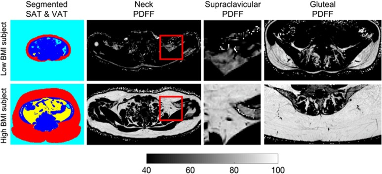
BACKGROUND/OBJECTIVES: The purpose of this study was to examine the relationship of the proton density fat fraction (PDFF), measured by magnetic resonance imaging (MRI), of supraclavicular and gluteal adipose tissue with subcutaneous and visceral adipose tissue (SAT and VAT) volumes, liver fat fraction and anthropometric obesity markers. The supraclavicular fossa was selected as a typical location where brown adipocytes may be present in humans and the gluteal region was selected as a typical location enclosing primarily white adipocytes.
SUBJECTS/METHODS: In this cross-sectional study, 61 adults (44 women, median age 29.3 years, range 21-68 years) underwent an MRI examination of the neck and the abdomen/pelvis (3T, Ingenia, Philips Healthcare). PDFF maps of the supraclavicular and gluteal adipose tissue and the liver were generated. Volumes of SAT and VAT were calculated and supraclavicular and subcutaneous fat were segmented using custom-built post-processing algorithms. Body mass index (BMI), waist circumference and waist-to-height ratio were recorded. Statistical analysis was conducted using the Student's t-test and Pearson correlation analysis.
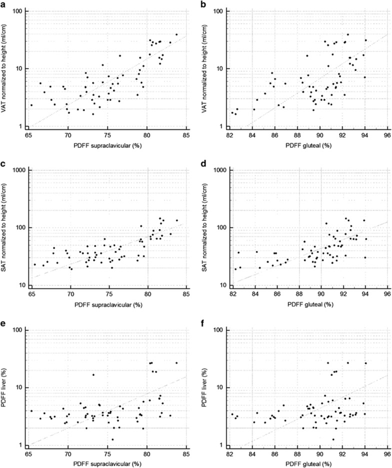
Mean supraclavicular PDFF was 75.3±4.7% (range 65.4-83.8%) and mean gluteal PDFF was 89.7±2.9% (range 82.2-94%), resulting in a significant difference (P<0.0001). Supraclavicular PDFF was positively correlated with VAT (r=0.76, P<0.0001), SAT (r=0.73, P<0.0001), liver PDFF (r=0.42, P=0.0008) and all measured anthropometric obesity markers. Gluteal subcutaneous PDFF also correlated with VAT (r=0.59, P<0.0001), SAT (r=0.63, P<0.0001), liver PDFF (r=0.3, P=0.02) and anthropometric obesity markers.
The positive correlations between adipose tissue PDFF and imaging, as well as anthropometric obesity markers suggest that adipose tissue PDFF may be useful as a biomarker for improving the characterization of the obese phenotype, for risk stratification and for selection of appropriate treatment strategies.
背景/目的:本研究旨在探讨磁共振成像(MRI)测量的锁骨上和臀区脂肪组织质子密度脂肪分数(PDFF)与皮下和内脏脂肪组织(SAT 和 VAT)体积、肝脂肪分数和人体肥胖标志物的关系。选择锁骨上窝作为可能存在棕色脂肪细胞的典型部位,选择臀区作为主要包含白色脂肪细胞的典型部位。
受试者/方法:在这项横断面研究中,61 名成年人(44 名女性,中位年龄 29.3 岁,范围 21-68 岁)接受了颈部和腹部/骨盆 MRI 检查(3T,Ingenia,飞利浦医疗保健)。生成锁骨上和臀区脂肪组织和肝脏的 PDFF 图。计算 SAT 和 VAT 的体积,并使用定制的后处理算法对锁骨上和皮下脂肪进行分割。记录体重指数(BMI)、腰围和腰高比。使用学生 t 检验和 Pearson 相关分析进行统计分析。
锁骨上 PDFF 的平均值为 75.3±4.7%(范围 65.4-83.8%),臀区 PDFF 的平均值为 89.7±2.9%(范围 82.2-94%),差异有统计学意义(P<0.0001)。锁骨上 PDFF 与 VAT(r=0.76,P<0.0001)、SAT(r=0.73,P<0.0001)、肝 PDFF(r=0.42,P=0.0008)和所有测量的人体肥胖标志物呈正相关。臀区皮下 PDFF 也与 VAT(r=0.59,P<0.0001)、SAT(r=0.63,P<0.0001)、肝 PDFF(r=0.3,P=0.02)和人体肥胖标志物呈正相关。
脂肪组织 PDFF 与影像学和人体肥胖标志物之间的正相关关系表明,脂肪组织 PDFF 可能作为一种生物标志物,有助于改善肥胖表型的特征描述、风险分层和选择合适的治疗策略。