Aaron Sanjith, Arthur Anupriya, Prabakhar A T, Mannam Pavitra, Shyamkumar N K, Mani Sunithi, Mathew Vivek, Peter Jeyanthi, Sivadasan Ajith, Alexander Anika, Karthik M, Benjamin Rohith Ninan, Alexander Mathew
Department of Neurological Sciences, Christian Medical College and Hospital, Vellore, Tamil Nadu, India.
Department of Ophthalmology, Christian Medical College and Hospital, Vellore, Tamil Nadu, India.
Ann Indian Acad Neurol. 2017 Jul-Sep;20(3):294-301. doi: 10.4103/aian.AIAN_11_17.
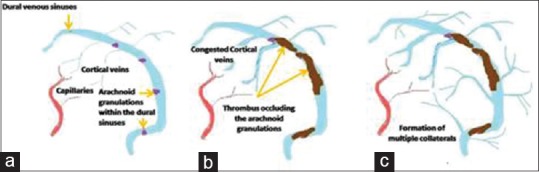
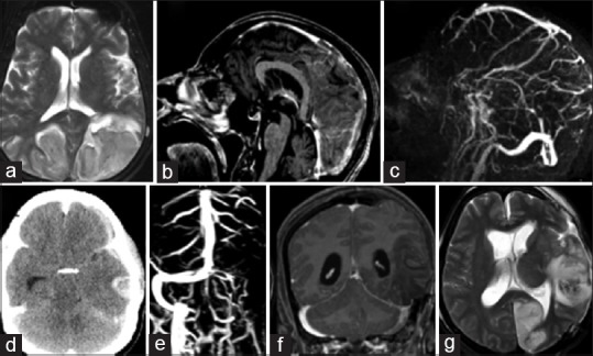
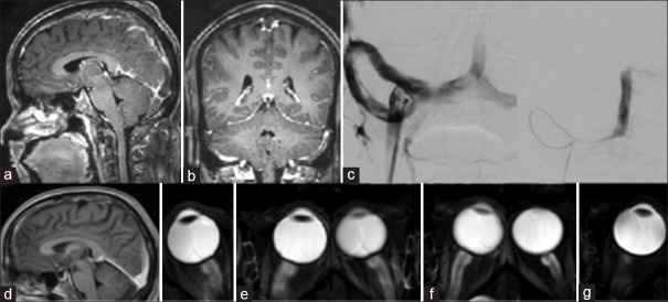
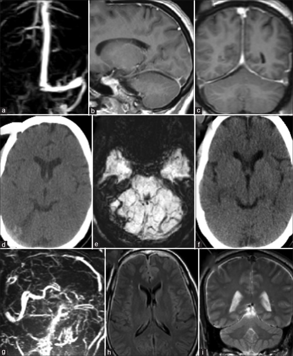
Visual impairment can complicate cerebral venous thrombosis (CVT). Here, we describe the various pathophysiological mechanisms and treatments available. A retrospective chart review of all patients treated for CVT in a large quaternary teaching hospital was done, and cases with visual impairment due to CVT were identified. The various mechanisms causing visual impairment in CVT were (1) raised intracranial pressure (ICP) caused by venous thrombosis without venous infarcts resulting in a benign intracranial hypertension-like presentation of CVT, (2) venous infarcts involving the occipital cortex, (3) raised ICP following the development of a secondary dural arteriovenous (AV) fistula, and (4) arterial occipital infarcts due to posterior cerebral artery compression secondary to herniation in large venous infarcts. Apart from using systemic anticoagulants to attempt recanalization and drugs with carbonic anhydrase inhibitor activity to reduce the ICPs, treatment modalities employed to save vision were (1) recanalization by local thrombolysis, stenting, or mechanical devices; (2) cerebrospinal fluid diversion procedures such as theco-periotoneal shunting; (3) optic nerve sheath fenestration; and (4) specific treatment for conditions such as dural AV fistula occurring as a late complication. CVT can cause visual impairment through different pathophysiological mechanisms. Depending on the mechanism, treatment strategies need to be tailored. Furthermore, very close monitoring is needed both in the acute and in the follow-up period, as new pathophysiological mechanisms can arise, compromising the vision. This may require a different treatment approach. Literature on this aspect of CVT is lacking.
视力障碍会使脑静脉血栓形成(CVT)复杂化。在此,我们描述各种可用的病理生理机制和治疗方法。我们对一家大型四级教学医院中所有接受CVT治疗的患者进行了回顾性病历审查,并确定了因CVT导致视力障碍的病例。CVT导致视力障碍的各种机制包括:(1)静脉血栓形成导致颅内压(ICP)升高,但无静脉梗死,导致CVT呈现类似良性颅内高压的表现;(2)累及枕叶皮质的静脉梗死;(3)继发硬脑膜动静脉(AV)瘘后ICP升高;(4)在大面积静脉梗死中,由于脑疝导致大脑后动脉受压引起的枕叶动脉梗死。除了使用全身抗凝剂试图实现再通以及使用具有碳酸酐酶抑制剂活性的药物降低ICP外,为挽救视力所采用的治疗方式包括:(1)通过局部溶栓、支架置入或机械装置进行再通;(2)脑脊液分流手术,如脑室 - 腹腔分流术;(3)视神经鞘开窗术;(4)针对作为晚期并发症出现的硬脑膜AV瘘等情况的特异性治疗。CVT可通过不同的病理生理机制导致视力障碍。根据机制的不同,需要制定相应的治疗策略。此外,在急性期和随访期都需要密切监测,因为可能会出现新的病理生理机制,从而损害视力。这可能需要采用不同的治疗方法。关于CVT这方面的文献较为缺乏。