Balan Maria, Hope Aimee, Cassidy Joseph, McCullough Maureen, O'Brien Peter J
Veterinary Pathobiology Section, University College Dublin, School of Veterinary Medicine, Dublin, Ireland.
Veterinary Clinical Sciences Section, University College Dublin, School of Veterinary Medicine, Dublin, Ireland.
JFMS Open Rep. 2017 Sep 19;3(2):2055116917730180. doi: 10.1177/2055116917730180. eCollection 2017 Jul-Dec.
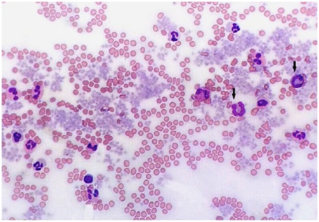
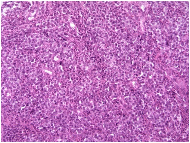
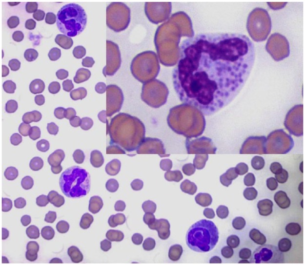
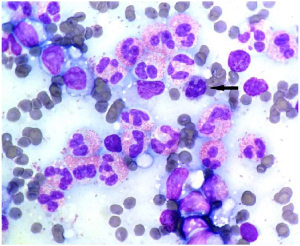
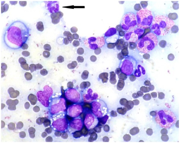
A 5-year-old male neutered domestic shorthair cat was referred with a history of persistent pyrexia, pica, soft faeces, inappetence, intermittent vomiting, mild-to-moderate granulocytosis and mild hypercalcaemia. No significant improvement was noted after antibiotic and corticosteroid treatment, except that the hypercalcaemia resolved. Physical examination, including thoracic auscultation, and abdominal and peripheral lymph node palpation, were unremarkable. On admission, haematology revealed moderate leukocytosis (36.8 × 10/l) with moderate-to-marked eosinophilia (21.3 × 10/l) and marked basophilia (4.04 × 10/l), the latter identified microscopically. Lymphocytes were markedly decreased (0.37 × 10/l). Blood smear examination revealed 58% eosinophils, 28% neutrophils, 11% basophils, 2% monocytes, 1% lymphocytes and marked, diffuse platelet clumping. Biochemistry abnormalities indicated mild pancreatitis, dehydration and anorexia with mildly increased pancreatic lipase, mild hypernatraemia (157 mmol/l), a moderate decrease in urea (3.1 mmol/l) and a slight decrease in phosphate (1.32 mmol/l). Ultrasound and radiographic imaging revealed enlargement of the mesenteric lymph nodes. Fine-needle aspiration, a Tru-cut biopsy and immunohistochemistry were performed. Cytological examination revealed 65-75% lymphocytes (80% were larger than a neutrophil), ~25-35% eosinophils and occasional basophils. Lymphocytes had single, small (<1/3 red blood cells), prominent nucleoli and increased pale, mildly vacuolated cytoplasm. On histopathology, cells were monomorphic, large, with prominent nucleoli, and mild, multifocal, staining for T-cell marker CD3. Smaller cells were strongly CD3-positive. Cells were negative for B-cell marker CD45R.
This is the most severe case of paraneoplastic basophilia reported with feline alimentary T-cell lymphoma with accompanying eosinophilia and lymph node infiltration. Feline basophil prevalence is reported for the first time.
一只5岁已绝育的雄性家猫因持续发热、异食癖、软便、食欲不振、间歇性呕吐、轻度至中度粒细胞增多症和轻度高钙血症前来就诊。抗生素和皮质类固醇治疗后未见明显改善,高钙血症除外。体格检查,包括胸部听诊、腹部及外周淋巴结触诊,均无异常。入院时,血液学检查显示中度白细胞增多(36.8×10⁹/L),伴有中度至重度嗜酸性粒细胞增多(21.3×10⁹/L)和重度嗜碱性粒细胞增多(4.04×10⁹/L),后者通过显微镜检查确定。淋巴细胞明显减少(0.37×10⁹/L)。血涂片检查显示嗜酸性粒细胞占58%,中性粒细胞占28%,嗜碱性粒细胞占11%,单核细胞占2%,淋巴细胞占1%,且有明显的弥漫性血小板聚集。生化异常表明有轻度胰腺炎、脱水和厌食,伴有胰脂肪酶轻度升高、轻度高钠血症(157 mmol/L)、尿素中度降低(3.1 mmol/L)和磷酸盐轻度降低(1.32 mmol/L)。超声和放射影像学检查显示肠系膜淋巴结肿大。进行了细针穿刺抽吸、Tru-cut活检和免疫组织化学检查。细胞学检查显示约65% - 75%为淋巴细胞(约80%大于中性粒细胞),约25% - 35%为嗜酸性粒细胞,偶见嗜碱性粒细胞。淋巴细胞有单个、小(<1/3红细胞)、明显的核仁,且苍白、轻度空泡化的细胞质增多。组织病理学检查显示细胞呈单形性、大,有明显核仁,轻度多灶性T细胞标志物CD3染色阳性。较小的细胞CD3强阳性。细胞B细胞标志物CD45R阴性。
这是报道的猫肠道T细胞淋巴瘤伴嗜酸性粒细胞增多和淋巴结浸润的最严重的副肿瘤性嗜碱性粒细胞增多症病例。首次报道了猫嗜碱性粒细胞的患病率。