Department of Medicine and Radiology, University of Chicago, Chicago, Illinois.
Departments of Medicine and Radiology and the Cardiovascular Imaging Center, University of Virginia Health System, Charlottesville, Virginia.
JACC Cardiovasc Imaging. 2017 Oct;10(10 Pt A):1180-1193. doi: 10.1016/j.jcmg.2017.08.005.
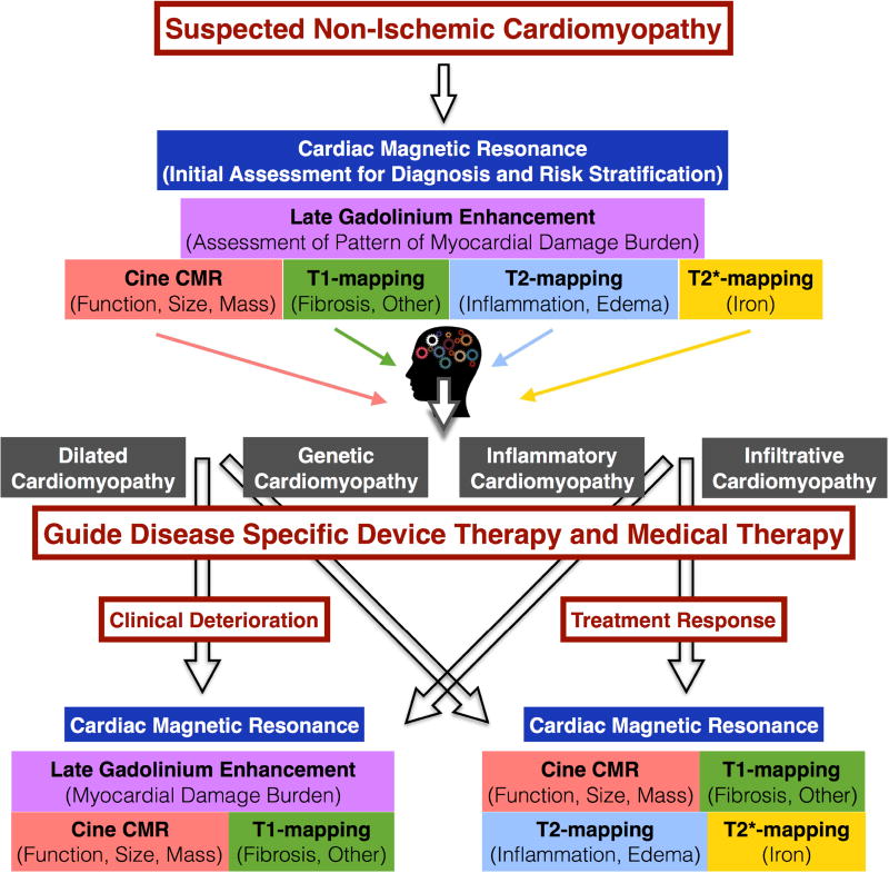
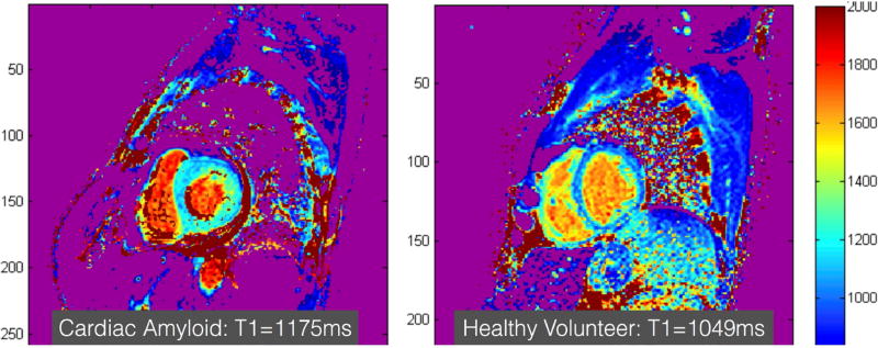
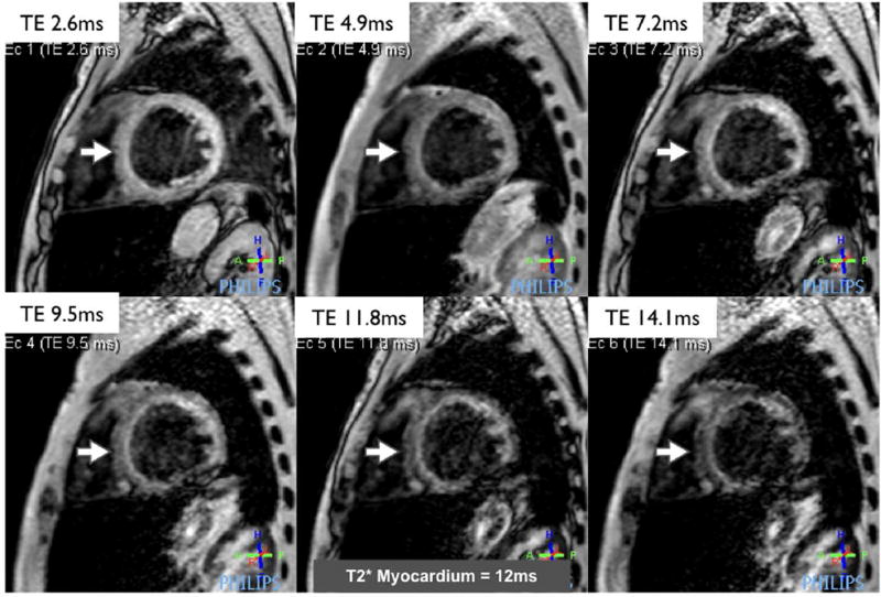
Cardiac magnetic resonance (CMR) is a valuable tool for the evaluation of patients with, or at risk for, heart failure and has a growing impact on diagnosis, clinical management, and decision making. Through its ability to characterize the myocardium by using multiple different imaging parameters, it provides insight into the etiology of the underlying heart failure and its prognosis. CMR is widely accepted as the reference standard for quantifying chamber size and ejection fraction. Additionally, tissue characterization techniques such as late gadolinium enhancement (LGE) and other quantitative parameters such as T mapping, both native and with measurement of extracellular volume fraction; T mapping; and T* mapping have been validated against histological findings in a wide range of clinical scenarios. In particular, the pattern of LGE in the myocardium can help determine the underlying etiology of the heart failure. The presence and extent of LGE determine prognosis in many of the nonischemic cardiomyopathies. The use of CMR should increase as its utility in characterization and assessment of prognosis in cardiomyopathies is increasingly recognized.
心脏磁共振(CMR)是评估心力衰竭患者或有心力衰竭风险患者的一种有价值的工具,它对诊断、临床管理和决策制定的影响越来越大。通过使用多种不同的成像参数来对心肌进行特征描述,CMR 可以深入了解心力衰竭的潜在病因及其预后。CMR 被广泛接受为量化心室大小和射血分数的参考标准。此外,组织特征描述技术,如延迟钆增强(LGE)和其他定量参数,如 T 映射,无论是原生的还是测量细胞外容积分数的;T 映射;以及 T*映射已经在广泛的临床情况下通过与组织学发现进行验证。特别是,心肌中 LGE 的模式可以帮助确定心力衰竭的潜在病因。在许多非缺血性心肌病中,LGE 的存在和程度决定了预后。随着 CMR 在心肌病的特征描述和预后评估中的应用越来越多,其使用也应增加。