Centro Nacional de Investigaciones Cardiovasculares Carlos III (CNIC) and Centro de Investigación Biomédica en Red de Enfermedades Respiratorias (CIBERES). C/Melchor Fernández-Almagro 3, 28029, Madrid, Spain.
Division of Hematopoietic Innovative Therapies, Centro de Investigaciones Energéticas Medioambientales y Tecnológicas/Centro de Investigación Biomédica en Red de Enfermedades Raras, 28040, Madrid, Spain.
Sci Rep. 2017 Oct 16;7(1):13242. doi: 10.1038/s41598-017-12829-y.
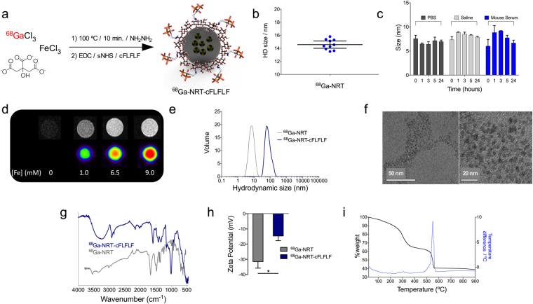
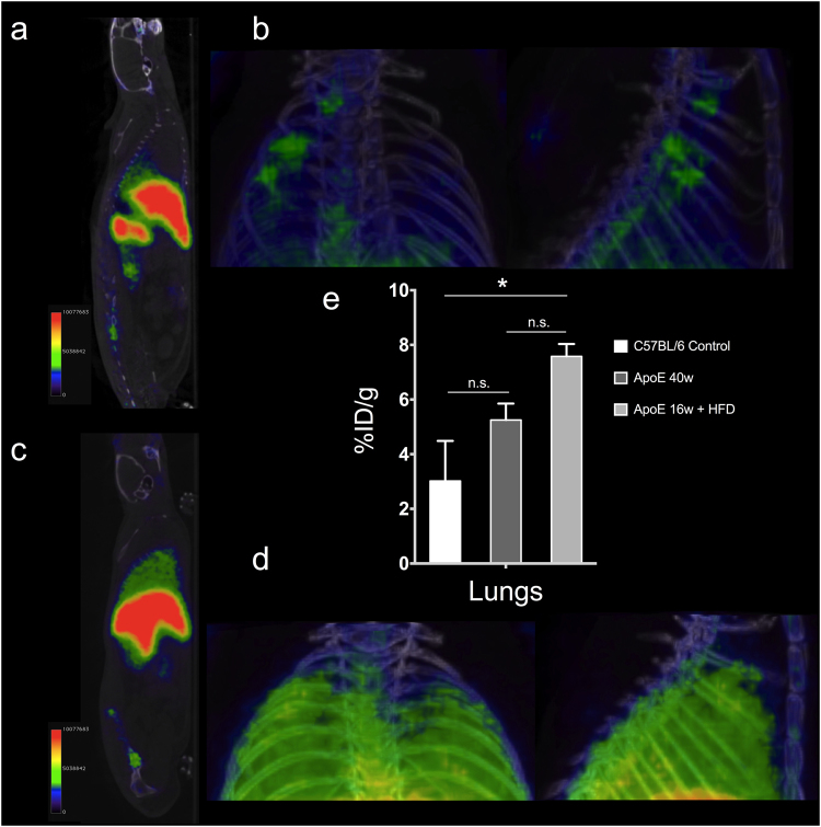
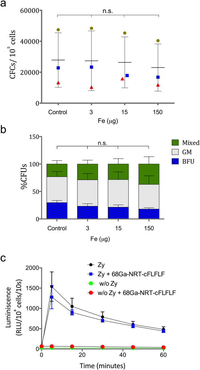
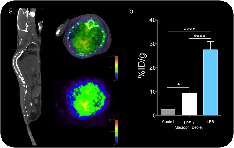
In vivo detection and quantification of inflammation is a major goal in molecular imaging. Furthermore, cell-specific detection of inflammation would be a tremendous advantage in the characterization of many diseases. Here, we show how this goal can be achieved through the synergistic combination of nanotechnology and nuclear imaging. One of the most remarkable features of this hybrid approach is the possibility to tailor the pharmacokinetics of the nanomaterial-incorporated biomolecule and radionuclide. A good example of this approach is the covalent binding of a large amount of a neutrophil-specific, hydrophobic peptide on the surface of Ga core-doped nanoparticles. This new nano-radiotracer has been used for non-invasive in vivo detection of acute inflammation with very high in vivo labelling efficiency, i.e. a large percentage of labelled neutrophils. Furthermore, we demonstrate that the tracer is neutrophil-specific and yields images of neutrophil recruitment of unprecedented quality. Finally, the nano-radiotracer was successfully detected in chronic inflammation in atherosclerosis-prone ApoE mice after several weeks on a high-fat diet.
在体检测和量化炎症是分子成像的主要目标。此外,炎症的细胞特异性检测将是许多疾病特征描述的巨大优势。在这里,我们展示了如何通过纳米技术和核医学成像的协同组合来实现这一目标。这种混合方法最显著的特点之一是能够调整纳米材料结合生物分子和放射性核素的药代动力学。这种方法的一个很好的例子是将大量嗜中性粒细胞特异性的疏水性肽共价结合到 Ga 核掺杂纳米颗粒的表面。这种新型纳米放射性示踪剂已用于急性炎症的非侵入性体内检测,具有非常高的体内标记效率,即标记的嗜中性粒细胞的百分比很大。此外,我们证明该示踪剂具有嗜中性粒细胞特异性,并产生了前所未有的质量的嗜中性粒细胞募集图像。最后,在高脂饮食数周后,在动脉粥样硬化易感 ApoE 小鼠的慢性炎症中成功检测到了纳米放射性示踪剂。