Li Hui, Zhao Jianmin
Department of Neurology, Xinxiang Central Hospital, Xinxiang, China.
Onco Targets Ther. 2017 Oct 6;10:4895-4904. doi: 10.2147/OTT.S141008. eCollection 2017.
has been indicated to act as a tumor suppressor in various cancers. However, the function and molecular mechanism of in meningioma progression have not been elucidated.
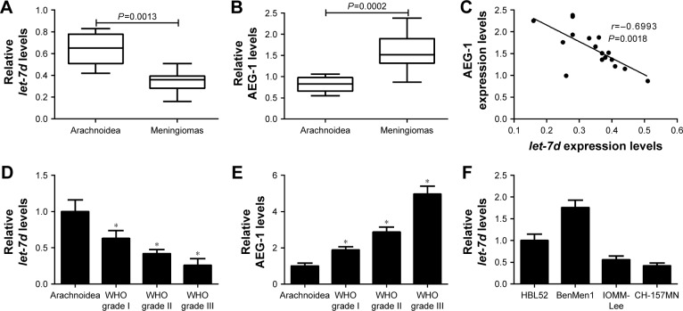
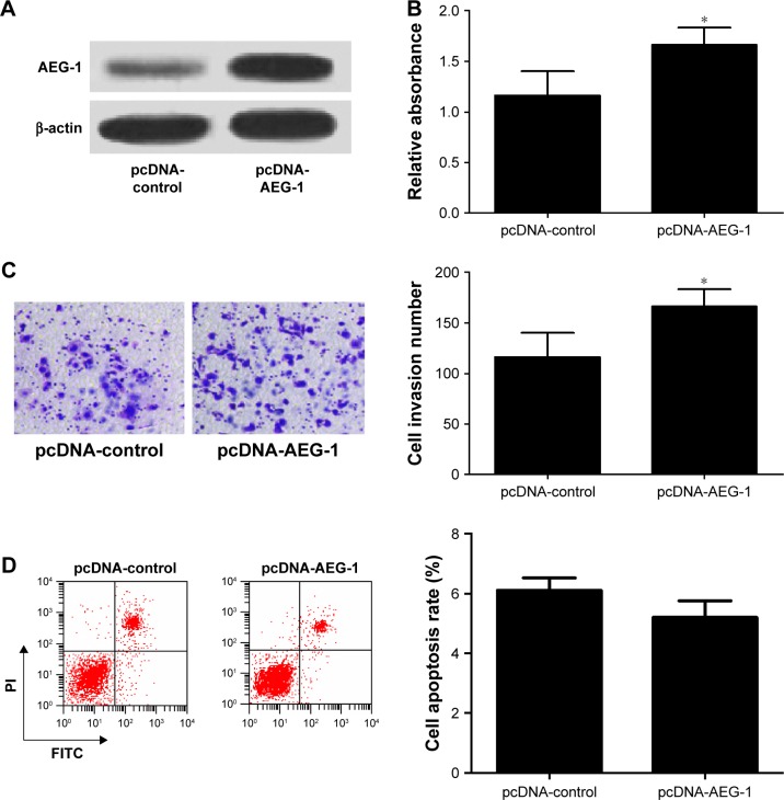
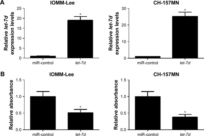
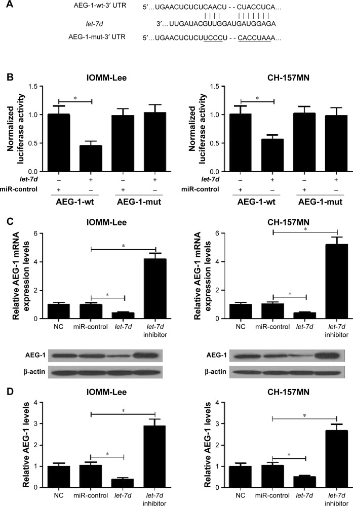
Quantitative real-time polymerase chain reaction (qRT-PCR) was performed to detect the expression levels of and AEG-1 mRNA in meningioma tissues and cell lines. The protein level of AEG-1 was measured by Western blot analysis. MTT assay, Transwell invasion assay and flow cytometry analysis were carried out to determine the proliferation, invasion and apoptosis of IOMM-Lee and CH-157MN cells, respectively. Target gene of was verified by luciferase reporter analysis.
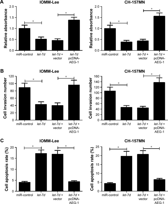
expression was downregulated, and AEG-1 expression was upregulated in meningioma tumor tissues. overexpression suppressed proliferation and invasion and induced apoptosis in IOMM-Lee and CH-157MN cells. Moreover, AEG-1 was a direct target of . Restoration of AEG-1 expression reversed -mediated suppression of the proliferation and invasion and -induced apoptosis in IOMM-Lee and CH-157MN cells.
repressed proliferation and invasion and promoted apoptosis of meningioma cells by targeting AEG-1. The present study provided a better understanding of the meningioma pathogenesis and a promising therapeutic target for meningioma patients.
已表明在多种癌症中发挥肿瘤抑制作用。然而,其在脑膜瘤进展中的功能和分子机制尚未阐明。
采用定量实时聚合酶链反应(qRT-PCR)检测脑膜瘤组织和细胞系中 及AEG-1 mRNA的表达水平。通过蛋白质印迹分析测定AEG-1的蛋白水平。分别进行MTT试验、Transwell侵袭试验和流式细胞术分析,以确定IOMM-Lee和CH-157MN细胞的增殖、侵袭和凋亡情况。通过荧光素酶报告基因分析验证 的靶基因。
在脑膜瘤肿瘤组织中, 表达下调,而AEG-1表达上调。 在IOMM-Lee和CH-157MN细胞中过表达可抑制增殖和侵袭并诱导凋亡。此外,AEG-1是 的直接靶标。恢复AEG-1表达可逆转 在IOMM-Lee和CH-157MN细胞中介导的对增殖和侵袭的抑制以及诱导的凋亡。
通过靶向AEG-1抑制脑膜瘤细胞的增殖和侵袭并促进其凋亡。本研究为更好地理解脑膜瘤发病机制以及为脑膜瘤患者提供了一个有前景的治疗靶点。