Flor Amy C, Wolfgeher Don, Wu Ding, Kron Stephen J
Department of Molecular Genetics and Cell Biology and Ludwig Center for Metastasis Research, The University of Chicago, Chicago, IL, USA.
Cell Death Discov. 2017 Oct 30;3:17075. doi: 10.1038/cddiscovery.2017.75. eCollection 2017.
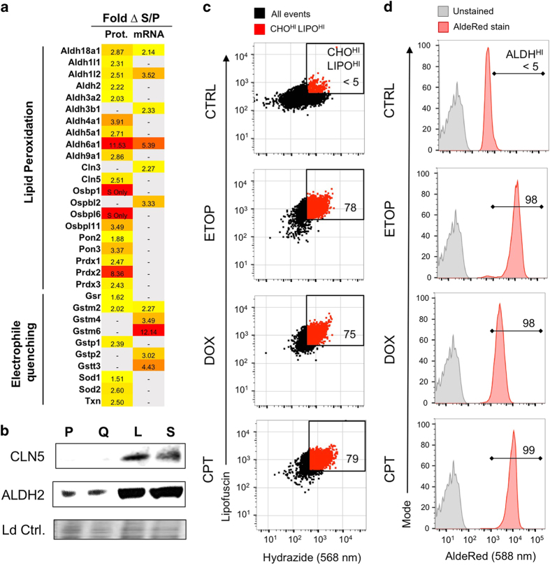
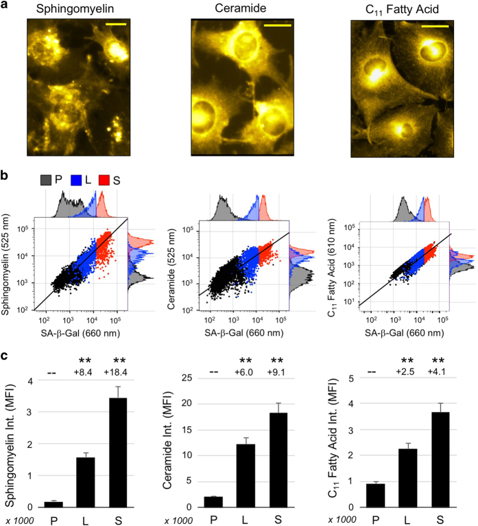
At their proliferative limit, normal cells arrest and undergo replicative senescence, displaying large cell size, flat morphology, and senescence-associated beta-galactosidase (SA--Gal) activity. Normal or tumor cells exposed to genotoxic stress undergo therapy-induced senescence (TIS), displaying a similar phenotype. Senescence is considered a DNA damage response, but cellular heterogeneity has frustrated identification of senescence-specific markers and targets. To explore the senescent cell proteome, we treated tumor cells with etoposide and enriched SA--Gal cells by fluorescence-activated cell sorting (FACS). The enriched TIS cells were compared to proliferating or quiescent cells by label-free quantitative LC-MS/MS proteomics and systems analysis, revealing activation of multiple lipid metabolism pathways. Senescent cells accumulated lipid droplets and imported lipid tracers, while treating proliferating cells with specific lipids induced senescence. Senescent cells also displayed increased lipid aldehydes and upregulation of aldehyde detoxifying enzymes. These results place deregulation of lipid metabolism alongside genotoxic stress as factors regulating cellular senescence.
在其增殖极限时,正常细胞会停滞并进入复制性衰老,表现出细胞体积大、形态扁平以及衰老相关β-半乳糖苷酶(SA-β-Gal)活性。暴露于基因毒性应激的正常细胞或肿瘤细胞会经历治疗诱导的衰老(TIS),表现出类似的表型。衰老被认为是一种DNA损伤反应,但细胞异质性阻碍了衰老特异性标志物和靶点的鉴定。为了探索衰老细胞蛋白质组,我们用依托泊苷处理肿瘤细胞,并通过荧光激活细胞分选(FACS)富集SA-β-Gal细胞。通过无标记定量液相色谱-质谱/质谱蛋白质组学和系统分析,将富集的TIS细胞与增殖或静止细胞进行比较,揭示了多种脂质代谢途径的激活。衰老细胞积累脂滴并摄取脂质示踪剂,而用特定脂质处理增殖细胞会诱导衰老。衰老细胞还表现出脂质醛增加以及醛解毒酶上调。这些结果表明脂质代谢失调与基因毒性应激一样,都是调节细胞衰老的因素。