Sonmez Kenan, Ozcan Pehmen Y
Ulucanlar Eye Training and Research Hospital, Ankara, Turkey.
Sanliurfa Education and Research Hospital, Sanliurfa, Turkey.
Case Rep Ophthalmol Med. 2017;2017:4630187. doi: 10.1155/2017/4630187. Epub 2017 Oct 4.
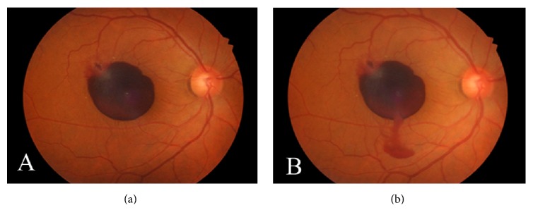
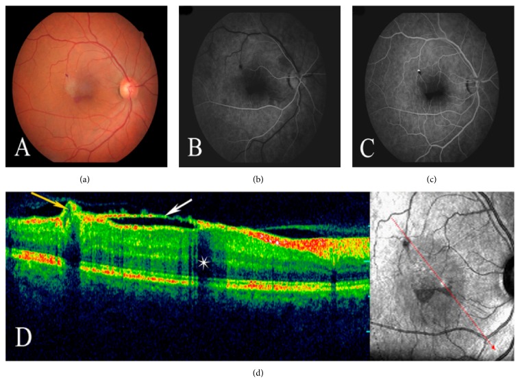
A 55-year-old man presented with sudden deterioration of vision in the right eye. His visual acuity was reduced to hand motion because of a large multilevel premacular hemorrhage. Nd:YAG laser was performed to drain the entrapped hemorrhage under the internal limiting membrane (ILM) and posterior hyaloid face in the macula into the vitreous. Immediately after laser treatment, streaming of red blood cells into the vitreous gel through the perforation site was observed. At the first-month follow-up, BCVA improved to 20/25 and ILM wrinkling was observed at the macula where the preretinal hemorrhage cleared. Fluorescein angiography revealed an isolated retinal venous macroaneurysm located on the macular branch of the superotemporal vein at the bifurcation site. In contrast to retinal arterial macroaneurysms, retinal venous macroaneurysms are quite rare. To the best of our knowledge, this is the first case reported with multilevel premacular hemorrhage caused by an isolated retinal venous macroaneurysm.
一名55岁男性因右眼视力突然下降前来就诊。由于黄斑前出现大量多层出血,其视力降至仅能感知手动。采用Nd:YAG激光将黄斑区内部限制膜(ILM)和后玻璃体膜下被困的出血引流至玻璃体。激光治疗后立即观察到红细胞通过穿孔部位流入玻璃体凝胶。在第一个月的随访中,最佳矫正视力(BCVA)提高到20/25,在黄斑区观察到ILM皱缩,此处视网膜前出血已清除。荧光素血管造影显示在颞上静脉黄斑分支的分叉处有一个孤立的视网膜静脉大动脉瘤。与视网膜动脉大动脉瘤相比,视网膜静脉大动脉瘤相当罕见。据我们所知,这是首例由孤立的视网膜静脉大动脉瘤引起多层黄斑前出血的病例报告。