Wang Xixi, Li Junyi, Yuan Yongsheng, Wang Min, Ding Jian, Zhang Jiejin, Zhu Lin, Shen Yuting, Zhang Hui, Zhang Kezhong
Department of Neurology, The First Affiliated Hospital of Nanjing Medical University, Nanjing, China.
Department of Radiology, The First Affiliated Hospital of Nanjing Medical University, Nanjing, China.
Oncotarget. 2017 Jul 5;8(46):81377-81386. doi: 10.18632/oncotarget.18996. eCollection 2017 Oct 6.
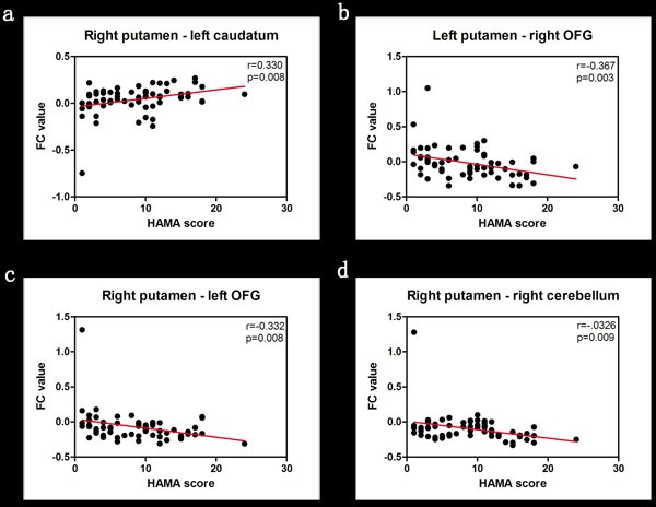
In this study, we used resting state-functional magnetic resonance imaging (rs-fMRI) to explore altered putamen functional connectivity (FC) in Parkinson's disease patients with anxiety disorder. We divided 65 Parkinson's disease patients into anxiety (PD-A; =18) and non-anxiety (PD-NA; =45) groups based on a Hamilton Anxiety Rating Scale cutoff score of 12. The PD-A patients exhibited altered putamen FC with cortical and subcortical regions. The PD-A patients showed enhanced putamen FC with the caudatum, which correlated with increased emotional processing during anxiety. Decreased putamen FC with the orbitofrontal gyrus and cerebellum also correlated with increased anxiety in Parkinson's disease. Our findings demonstrate that anxiety disorder in Parkinson's disease is associated with abnormal putamen FC networks, especially with caudatum, orbitofrontal gyrus and cerebellum.
在本研究中,我们使用静息态功能磁共振成像(rs-fMRI)来探究伴有焦虑症的帕金森病患者壳核功能连接(FC)的改变。我们根据汉密尔顿焦虑量表12分的临界值,将65例帕金森病患者分为焦虑组(PD-A;n = 18)和非焦虑组(PD-NA;n = 45)。PD-A患者的壳核与皮质及皮质下区域的FC发生了改变。PD-A患者壳核与尾状核的FC增强,这与焦虑期间情绪加工增加相关。壳核与眶额回及小脑的FC降低也与帕金森病患者焦虑增加相关。我们的研究结果表明,帕金森病中的焦虑症与异常的壳核FC网络有关,尤其是与尾状核、眶额回和小脑有关。