Barua Nabanita, Sitaraman Chitra, Goel Sonu, Chakraborti Chandana, Mukherjee Sonai, Parashar Hemandra
Department of Ophthalmology, Calcutta National Medical College, Kolkata, West Bengal, India.
Department of Ophthalmology, Anand Hospital and Eye Centre, Jaipur, Rajasthan, India.
Indian J Ophthalmol. 2016 Apr;64(4):296-302. doi: 10.4103/0301-4738.182941.
Analysis of diagnostic ability of macular ganglionic cell complex and retinal nerve fiber layer (RNFL) in glaucoma.
To correlate functional and structural parameters and comparing predictive value of each of the structural parameters using Fourier-domain (FD) optical coherence tomography (OCT) among primary open angle glaucoma (POAG) and ocular hypertension (OHT) versus normal population.
Single centric, cross-sectional study done in 234 eyes.
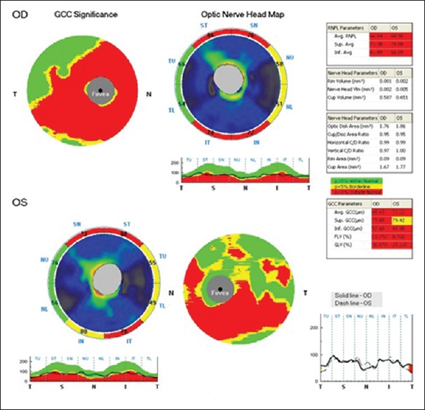
Patients were enrolled in three groups: POAG, ocular hypertensive and normal (40 patients in each group). After comprehensive ophthalmological examination, patients underwent standard automated perimetry and FD-OCT scan in optic nerve head and ganglion cell mode. The relationship was assessed by correlating ganglion cell complex (GCC) parameters with mean deviation. Results were compared with RNFL parameters.
Data were analyzed with SPSS, analysis of variance, t-test, Pearson's coefficient, and receiver operating curve.
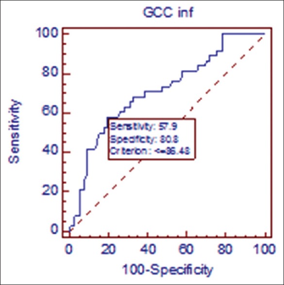
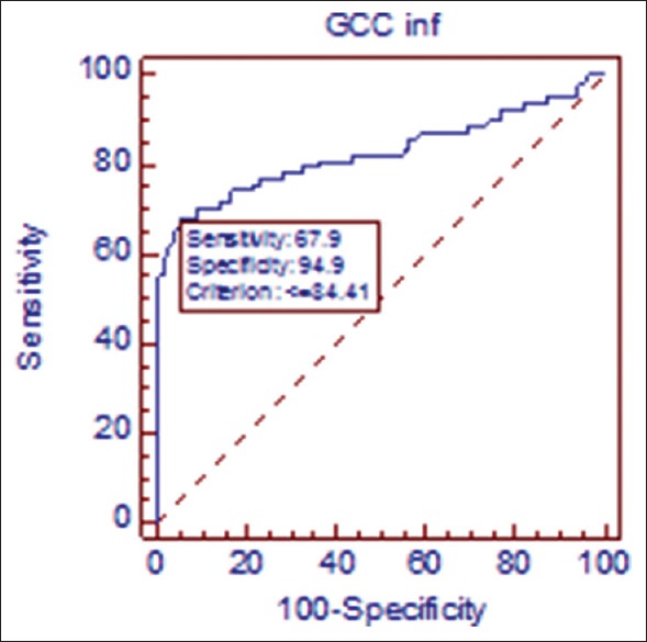
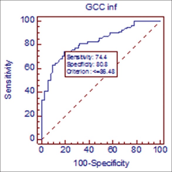
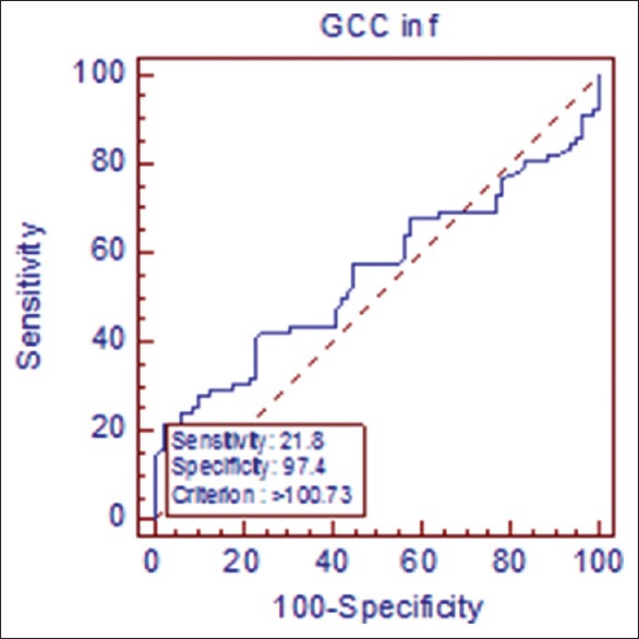
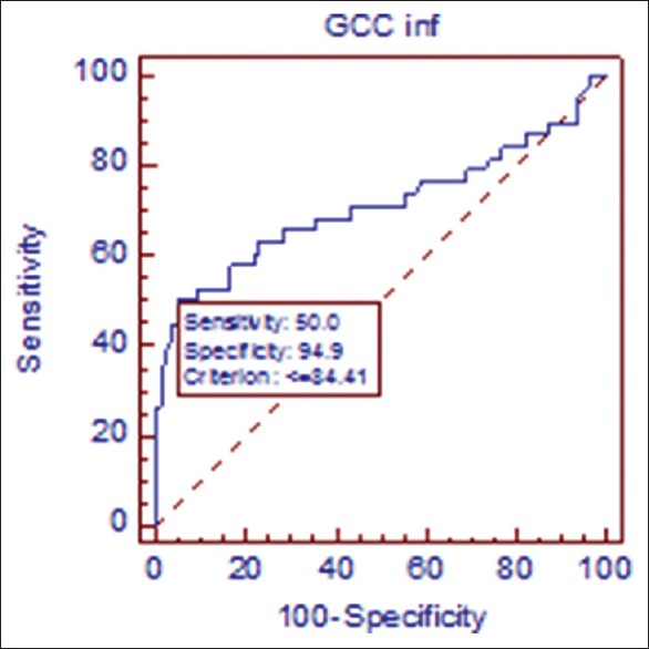
All parameters showed strong correlation with visual field (P < 0.001). Inferior GCC had highest area under curve (AUC) for detecting glaucoma (0.827) in POAG from normal population. However, the difference was not statistically significant (P > 0.5) when compared with other parameters. None of the parameters showed significant diagnostic capability to detect OHT from normal population. In diagnosing early glaucoma from OHT and normal population, only inferior GCC had statistically significant AUC value (0.715).
In this study, GCC and RNFL parameters showed equal predictive capability in perimetric versus normal group. In early stage, inferior GCC was the best parameter. In OHT population, single day cross-sectional imaging was not valuable.
青光眼黄斑神经节细胞复合体和视网膜神经纤维层(RNFL)诊断能力的分析。
关联功能和结构参数,并比较使用傅里叶域(FD)光学相干断层扫描(OCT)检测原发性开角型青光眼(POAG)和高眼压症(OHT)与正常人群时各结构参数的预测价值。
在234只眼中进行的单中心横断面研究。
患者分为三组:POAG组、高眼压组和正常组(每组40例患者)。经过全面的眼科检查后,患者接受标准自动视野检查以及视神经乳头和神经节细胞模式的FD-OCT扫描。通过将神经节细胞复合体(GCC)参数与平均偏差进行关联来评估两者之间的关系。结果与RNFL参数进行比较。
使用SPSS、方差分析、t检验、Pearson系数和受试者工作特征曲线对数据进行分析。
所有参数均与视野显示出强相关性(P < 0.001)。在POAG组中,下方GCC检测青光眼的曲线下面积(AUC)最高(0.827),用于区分正常人群。然而,与其他参数相比,差异无统计学意义(P > 0.5)。没有参数显示出从正常人群中检测OHT的显著诊断能力。在从OHT和正常人群中诊断早期青光眼时,只有下方GCC具有统计学意义的AUC值(0.715)。
在本研究中,GCC和RNFL参数在视野与正常组中显示出相同的预测能力。在早期阶段,下方GCC是最佳参数。在OHT人群中,单日横断面成像没有价值。