Department of Psychiatry and Psychotherapy, Medical University of Vienna, Waehringer Guertel 18-20, 1090, Vienna, Austria.
Division of Nuclear Medicine, Department of Biomedical Imaging and Image-guided Therapy, Medical University of Vienna, Vienna, Austria.
Brain Struct Funct. 2018 Apr;223(3):1369-1378. doi: 10.1007/s00429-017-1558-0. Epub 2017 Nov 13.
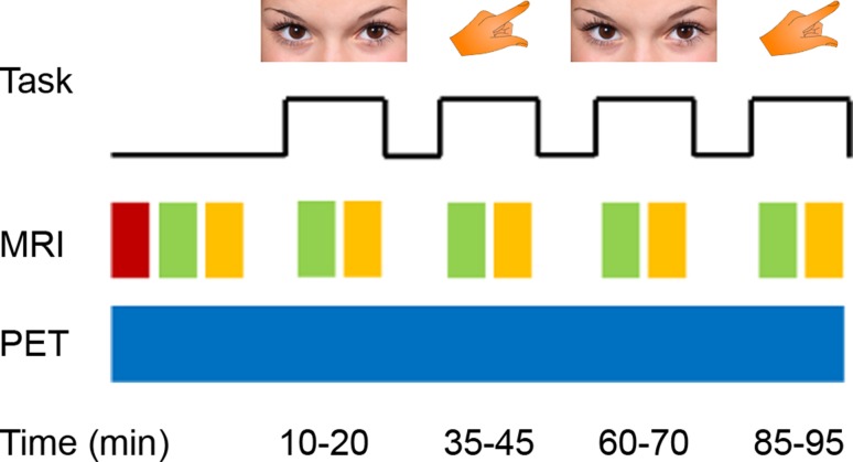
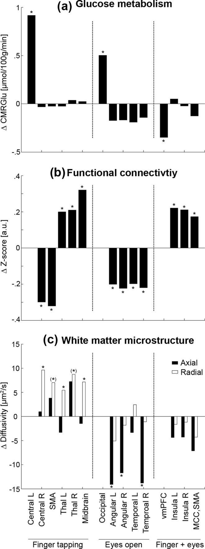
Except for task-specific functional MRI, the vast majority of imaging studies assessed human brain function at resting conditions. However, tracking task-specific neuronal activity yields important insight how the brain responds to stimulation. We specifically investigated changes in glucose metabolism, functional connectivity and white matter microstructure during task performance using several recent methodological advancements. Opening the eyes and right finger tapping had elicited an increased glucose metabolism in primary visual and motor cortices, respectively. Furthermore, a decreased metabolism was observed in the regions of the default mode network, which allowed absolute quantification of commonly described deactivations during cognitive tasks. These brain regions showed widespread task-specific changes in functional connectivity, which stretched beyond their primary resting-state networks and presumably reflected the level of recruitment of certain brain regions for each task. Finally, the corresponding white matter fiber pathways exhibited changes in axial and radial diffusivity during the tasks, which were regionally distinctive for certain tract groups. These results highlight that even simple task performance leads to substantial changes of entire brain networks. Exploiting the complementary nature of the different imaging modalities may reveal novel insights how the brain processes external stimuli and which networks are involved in certain tasks.
除了特定任务的功能磁共振成像,绝大多数成像研究都在静息状态下评估人脑功能。然而,追踪特定任务的神经元活动可以深入了解大脑如何对刺激做出反应。我们使用了几种最新的方法学进展,专门研究了任务执行过程中葡萄糖代谢、功能连接和白质微观结构的变化。睁开眼睛和用右手手指敲击分别引起初级视觉和运动皮层的葡萄糖代谢增加。此外,在默认模式网络的区域观察到代谢减少,这允许对认知任务期间通常描述的去激活进行绝对定量。这些脑区表现出广泛的特定任务功能连接变化,超出了它们的主要静息状态网络,并可能反映了特定脑区对每个任务的招募水平。最后,相应的白质纤维通路在任务期间表现出轴向和径向扩散率的变化,对于某些束组而言,这些变化在区域上是独特的。这些结果强调,即使是简单的任务执行也会导致整个大脑网络的实质性变化。利用不同成像模式的互补性质可能会揭示大脑如何处理外部刺激以及哪些网络参与特定任务的新见解。