Guangdong Provincial Key Laboratory of Medical Image Processing, School of Biomedical Engineering, Southern Medical University, Guangzhou 510515, China.
Center for Biostatistics, The Ohio State University Wexner Medical Center.
Bioinformatics. 2018 Mar 15;34(6):1024-1030. doi: 10.1093/bioinformatics/btx723.
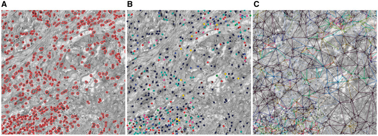
As a highly heterogeneous disease, the progression of tumor is not only achieved by unlimited growth of the tumor cells, but also supported, stimulated, and nurtured by the microenvironment around it. However, traditional qualitative and/or semi-quantitative parameters obtained by pathologist's visual examination have very limited capability to capture this interaction between tumor and its microenvironment. With the advent of digital pathology, computerized image analysis may provide a better tumor characterization and give new insights into this problem.
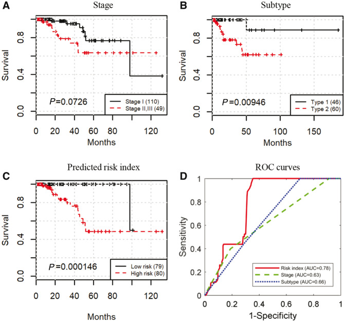
We propose a novel bioimage informatics pipeline for automatically characterizing the topological organization of different cell patterns in the tumor microenvironment. We apply this pipeline to the only publicly available large histopathology image dataset for a cohort of 190 patients with papillary renal cell carcinoma obtained from The Cancer Genome Atlas project. Experimental results show that the proposed topological features can successfully stratify early- and middle-stage patients with distinct survival, and show superior performance to traditional clinical features and cellular morphological and intensity features. The proposed features not only provide new insights into the topological organizations of cancers, but also can be integrated with genomic data in future studies to develop new integrative biomarkers.
https://github.com/chengjun583/KIRP-topological-features.
1271992826@qq.com or kunhuang@iu.edu.
Supplementary data are available at Bioinformatics online.
作为一种高度异质的疾病,肿瘤的进展不仅是通过肿瘤细胞的无限生长来实现的,还得到了其周围微环境的支持、刺激和滋养。然而,病理学家通过视觉检查获得的传统定性和/或半定量参数,其捕捉肿瘤与其微环境之间相互作用的能力非常有限。随着数字病理学的出现,计算机图像分析可能提供更好的肿瘤特征描述,并为解决这一问题提供新的见解。
我们提出了一种新的生物图像信息学管道,用于自动描述肿瘤微环境中不同细胞模式的拓扑组织。我们将该管道应用于唯一公开的、来自癌症基因组图谱项目的 190 名乳头状肾细胞癌患者的大型组织病理学图像数据集,以应用于该数据集。实验结果表明,所提出的拓扑特征可以成功地对早期和中期具有不同生存情况的患者进行分层,并且比传统的临床特征、细胞形态和强度特征具有更好的性能。所提出的特征不仅为癌症的拓扑组织提供了新的见解,而且还可以与未来研究中的基因组数据集成,以开发新的综合生物标志物。
https://github.com/chengjun583/KIRP-topological-features。
1271992826@qq.com 或 kunhuang@iu.edu。
补充数据可在生物信息学在线获得。