Department of Radiology and Nuclear Medicine, Radboud University Medical Center, Geert Grooteplein 10, 6525GA, Nijmegen, The Netherlands.
Department of Neurology, Radboud University Medical Center, Geert Grooteplein 10, 6525GA, Nijmegen, The Netherlands.
Sci Rep. 2017 Nov 15;7(1):15622. doi: 10.1038/s41598-017-15617-w.
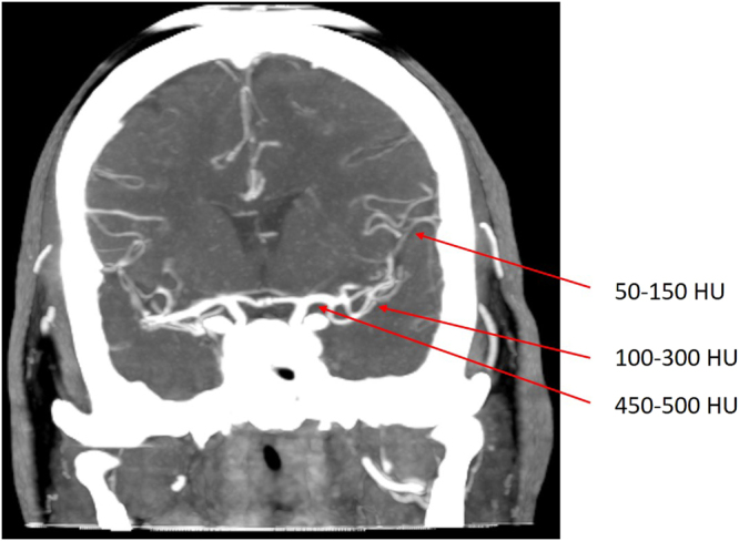
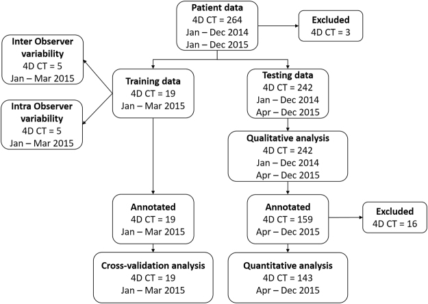
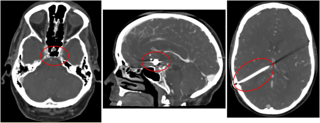
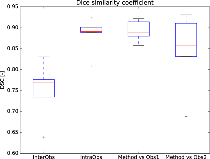
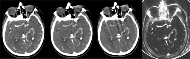
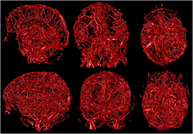
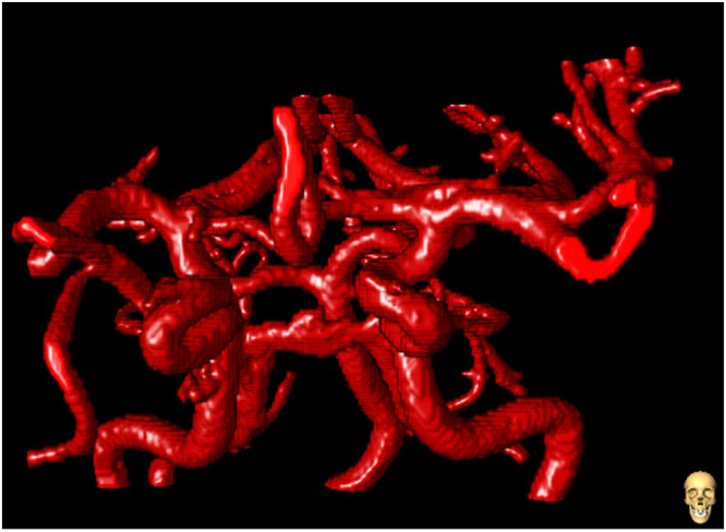
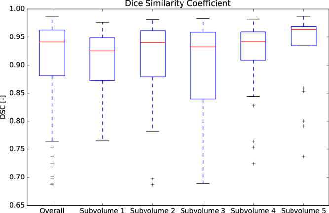
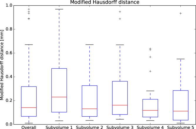
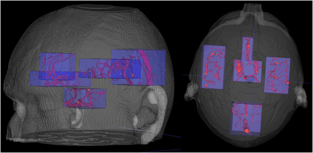
A robust method is presented for the segmentation of the full cerebral vasculature in 4-dimensional (4D) computed tomography (CT). The method consists of candidate vessel selection, feature extraction, random forest classification and postprocessing. Image features include among others the weighted temporal variance image and parameters, including entropy, of an intensity histogram in a local region at different scales. These histogram parameters revealed to be a strong feature in the detection of vessels regardless of shape and size. The method was trained and tested on a large database of 264 patients with suspicion of acute ischemia who underwent 4D CT in our hospital in the period January 2014 to December 2015. Five subvolumes representing different regions of the cerebral vasculature were annotated in each image in the training set by medical assistants. The evaluation was done on 242 patients. A total of 16 (<8%) patients showed severe under or over segmentation and were reported as failures. One out of five subvolumes was randomly annotated in 159 patients and was used for quantitative evaluation. Quantitative evaluation showed a Dice coefficient of 0.91 ± 0.07 and a modified Hausdorff distance of 0.23 ± 0.22 mm. Therefore, robust vessel segmentation in 4D CT is feasible with good accuracy.
提出了一种稳健的方法,用于对 4 维(4D)计算机断层扫描(CT)中的全脑血管进行分割。该方法包括候选血管选择、特征提取、随机森林分类和后处理。图像特征包括加权时间方差图像和局部区域不同尺度的强度直方图的熵等参数。这些直方图参数在检测血管时,无论形状和大小如何,都是一个很强的特征。该方法在我们医院于 2014 年 1 月至 2015 年 12 月期间对 264 名疑似急性缺血患者的大型 4D CT 数据库进行了训练和测试。在训练集中,由医疗助理对每张图像的五个子体积进行不同脑血管区域的注释。对 242 名患者进行了评估。共有 16 名(<8%)患者存在严重的分割不足或过度,被报告为失败。在 159 名患者中随机注释了一个子体积,并用于定量评估。定量评估显示,Dice 系数为 0.91±0.07,改进的 Hausdorff 距离为 0.23±0.22mm。因此,4D CT 中稳健的血管分割具有良好的准确性。