Department of Biomedical Engineering, University of Arizona, United States.
Department of Radiology, Stanford University, United States.
Neuroimage Clin. 2021;30:102573. doi: 10.1016/j.nicl.2021.102573. Epub 2021 Jan 26.
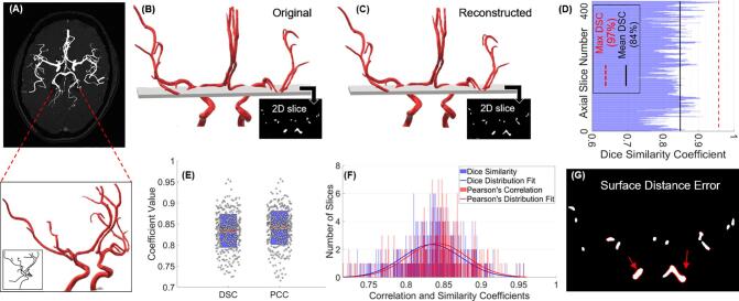
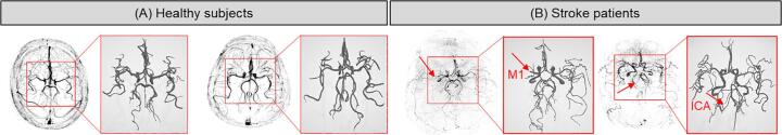
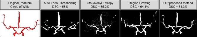
Accurate segmentation of cerebral vasculature and a quantitative assessment of its morphology is critical to various diagnostic and therapeutic purposes and is pertinent to studying brain health and disease. However, this is still a challenging task due to the complexity of the vascular imaging data. We propose an automated method for cerebral vascular segmentation without the need of any manual intervention as well as a method to skeletonize the binary segmented map to extract vascular geometric features and characterize vessel structure. We combine a Hessian-based probabilistic vessel-enhancing filtering with an active-contour-based technique to segment magnetic resonance and computed tomography angiograms (MRA and CTA) and subsequently extract the vessel centerlines and diameters to calculate the geometrical properties of the vasculature. Our method was validated using a 3D phantom of the Circle-of-Willis region, demonstrating 84% mean Dice similarity coefficient (DSC) and 85% mean Pearson's correlation coefficient (PCC) with minimal modified Hausdorff distance (MHD) error (3 surface pixels at most), and showed superior performance compared to existing segmentation algorithms upon quantitative comparison using DSC, PCC and MHD. We subsequently applied our algorithm to a dataset of 40 subjects, including 1) MRA scans of healthy subjects (n = 10, age = 30 ± 9), 2) MRA scans of stroke patients (n = 10, age = 51 ± 15), 3) CTA scans of healthy subjects (n = 10, age = 62 ± 12), and 4) CTA scans of stroke patients (n = 10, age = 68 ± 11), and obtained a quantitative comparison between the stroke and normal vasculature for both imaging modalities. The vascular network in stroke patients compared to age-adjusted healthy subjects was found to have a significantly (p < 0.05) higher tortuosity (3.24 ± 0.88 rad/cm vs. 7.17 ± 1.61 rad/cm for MRA, and 4.36 ± 1.32 rad/cm vs. 7.80 ± 0.92 rad/cm for CTA), higher fractal dimension (1.36 ± 0.28 vs. 1.71 ± 0.14 for MRA, and 1.56 ± 0.05 vs. 1.69 ± 0.20 for CTA), lower total length (3.46 ± 0.99 m vs. 2.20 ± 0.67 m for CTA), lower total volume (61.80 ± 18.79 ml vs. 34.43 ± 22.9 ml for CTA), lower average diameter (2.4 ± 0.21 mm vs. 2.18 ± 0.07 mm for CTA), and lower average branch length (4.81 ± 1.97 mm vs. 8.68 ± 2.03 mm for MRA), respectively. We additionally studied the change in vascular features with respect to aging and imaging modality. While we observed differences between features as a result of aging, statistical analysis did not show any significant differences, whereas we found that the number of branches were significantly different (p < 0.05) between the two imaging modalities (201 ± 73 for MRA vs. 189 ± 69 for CTA). Our segmentation and feature extraction algorithm can be applied on any imaging modality and can be used in the future to automatically obtain the 3D segmented vasculature for diagnosis and treatment planning as well as to study morphological changes due to stroke and other cerebrovascular diseases (CVD) in the clinic.
准确分割脑血管并对其形态进行定量评估对于各种诊断和治疗目的至关重要,并且与研究大脑健康和疾病有关。然而,由于血管成像数据的复杂性,这仍然是一项具有挑战性的任务。我们提出了一种无需任何手动干预即可自动分割脑血管的方法,以及一种将二值分割图骨架化以提取血管几何特征和描述血管结构的方法。我们结合了基于 Hessian 的概率血管增强滤波和基于主动轮廓的技术来分割磁共振和计算机断层血管造影(MRA 和 CTA),并随后提取血管中心线和直径以计算血管的几何性质。我们的方法使用 Circle-of-Willis 区域的 3D 体模进行了验证,证明了 84%的平均 Dice 相似系数(DSC)和 85%的平均 Pearson 相关系数(PCC),最小修正 Hausdorff 距离(MHD)误差(最多 3 个表面像素),并且在使用 DSC、PCC 和 MHD 进行定量比较时,与现有的分割算法相比表现出优越的性能。随后,我们将我们的算法应用于包括以下内容的 40 名受试者的数据集:1)健康受试者的 MRA 扫描(n=10,年龄=30±9),2)中风患者的 MRA 扫描(n=10,年龄=51±15),3)健康受试者的 CTA 扫描(n=10,年龄=62±12),和 4)中风患者的 CTA 扫描(n=10,年龄=68±11),并获得了两种成像方式中风和正常血管之间的定量比较。与年龄匹配的健康受试者相比,中风患者的血管网络被发现具有显著更高的迂曲度(3.24±0.88 rad/cm 与 7.17±1.61 rad/cm 用于 MRA,以及 4.36±1.32 rad/cm 与 7.80±0.92 rad/cm 用于 CTA)、更高的分形维数(1.36±0.28 与 1.71±0.14 用于 MRA,以及 1.56±0.05 与 1.69±0.20 用于 CTA)、更低的总长度(3.46±0.99 m 与 2.20±0.67 m 用于 CTA)、更低的总容积(61.80±18.79 ml 与 34.43±22.9 ml 用于 CTA)、更低的平均直径(2.4±0.21 mm 与 2.18±0.07 mm 用于 CTA)和更低的平均分支长度(4.81±1.97 mm 与 8.68±2.03 mm 用于 MRA)。我们还研究了血管特征随年龄和成像方式的变化。虽然我们观察到由于年龄而导致的特征差异,但统计分析没有显示出任何显著差异,而我们发现两种成像方式之间的分支数量存在显著差异(p<0.05)(201±73 用于 MRA 与 189±69 用于 CTA)。我们的分割和特征提取算法可应用于任何成像方式,并可用于未来自动获得用于诊断和治疗计划的 3D 分割血管,以及研究由于中风和其他脑血管疾病(CVD)引起的形态变化在临床实践中。