Choi Kyu Young, Cho Sung Woo, Choi Jun-Jae, Zhang Yu-Lian, Kim Dae Woo, Han Doo Hee, Kim Hyun Jik, Kim Dong-Young, Rhee Chae-Seo, Won Tae-Bin
Department of Otorhinolaryngology-Head and Neck Surgery, Hallym University College of Medicine, Kangnam Sacred Heart Hospital, Seoul, 07441, South Korea.
Department of Otorhinolaryngology-Head and Neck Surgery, Seoul National University College of Medicine, Seoul National University Hospital, Seoul, 03080, South Korea.
World J Otorhinolaryngol Head Neck Surg. 2017 Mar 9;3(1):17-23. doi: 10.1016/j.wjorl.2017.02.004. eCollection 2017 Mar.
The aim of this study was to investigate the regeneration process of the nasal mucosa after a surgically created mucosal defect in the rabbit nasal septum, and to evaluate the effects of different interventions.
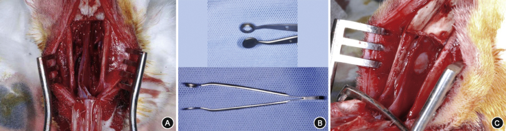
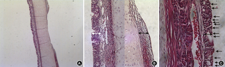
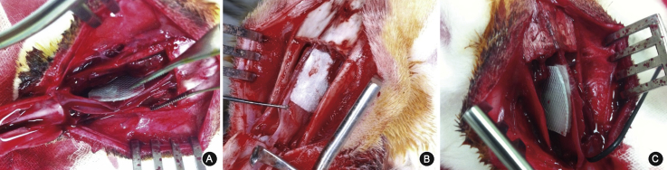
A 7 mm-diameter circular mucosal defect was made in the septum of forty New Zealand white rabbits. The rabbits were divided into four groups (ten rabbits in each group) according to the type of intervention; no treatment (control), silastic sheet (SS), hyaluronic acid (HA), and silastic sheet and hyaluronic acid (SS + HA) group. The diameter of the defect, mucosal thickness, epithelial thickness, and ciliated cell count were evaluated every week for five weeks.
The average diameter of the defect in the control group were 5.1, 3.65, 1.2, 0.75, and 0.05 mm at postoperative 1, 2, 3, 4, and 5 weeks. In the SS group, the diameter decreased to 4.35, 2.1, 0.35, 0.15, and 0 mm at postoperative 1, 2, 3, 4, and 5 weeks, respectively, in which the mean diameter of the postoperative week 2 was significantly smaller compared to control (3.65 mm vs. 2.1 mm, = 0.039). For the HA group and SS + HA group, the diameter of the defect did not show a significant difference from the control group during the five weeks. The mucosal thickness, epithelial thickness, and ciliated cell count of the regenerated mucosa were not significantly different among the groups.
The regeneration process of the nasal septal mucosa was identified using a novel rabbit model. Mucosal regeneration can be accelerated by applying silastic sheets.
本研究旨在探讨兔鼻中隔手术造成黏膜缺损后鼻黏膜的再生过程,并评估不同干预措施的效果。
在40只新西兰白兔的鼻中隔上制作直径7mm的圆形黏膜缺损。根据干预类型将兔子分为四组(每组10只):不治疗(对照组)、硅橡胶片(SS)组、透明质酸(HA)组和硅橡胶片加透明质酸(SS+HA)组。连续五周每周评估缺损直径、黏膜厚度、上皮厚度和纤毛细胞计数。
对照组术后第1、2、3、4和5周缺损的平均直径分别为5.1、3.65、1.2、0.75和0.05mm。在SS组中,术后第1、2、3、4和5周缺损直径分别降至4.35、2.1、0.35、0.15和0mm,其中术后第2周的平均直径与对照组相比显著更小(3.65mm对2.1mm,P=0.039)。对于HA组和SS+HA组,在五周内缺损直径与对照组相比无显著差异。再生黏膜的黏膜厚度、上皮厚度和纤毛细胞计数在各组之间无显著差异。
使用新型兔模型确定了鼻中隔黏膜的再生过程。应用硅橡胶片可加速黏膜再生。