• 文献检索
  • 文档翻译
  • 深度研究
  • 学术资讯
  • Suppr Zotero 插件Zotero 插件
  • 邀请有礼
  • 套餐&价格
  • 历史记录
应用&插件
Suppr Zotero 插件Zotero 插件浏览器插件Mac 客户端Windows 客户端微信小程序
定价
高级版会员购买积分包购买API积分包
服务
文献检索文档翻译深度研究API 文档MCP 服务
关于我们
关于 Suppr公司介绍联系我们用户协议隐私条款
关注我们

Suppr 超能文献

核心技术专利:CN118964589B侵权必究
粤ICP备2023148730 号-1Suppr @ 2026

文献检索

告别复杂PubMed语法,用中文像聊天一样搜索,搜遍4000万医学文献。AI智能推荐,让科研检索更轻松。

立即免费搜索

文件翻译

保留排版,准确专业,支持PDF/Word/PPT等文件格式,支持 12+语言互译。

免费翻译文档

深度研究

AI帮你快速写综述,25分钟生成高质量综述,智能提取关键信息,辅助科研写作。

立即免费体验

结直肠活检标本全切片图像的自动化组织学分类

Automated histological classification of whole slide images of colorectal biopsy specimens.

作者信息

Yoshida Hiroshi, Yamashita Yoshiko, Shimazu Taichi, Cosatto Eric, Kiyuna Tomoharu, Taniguchi Hirokazu, Sekine Shigeki, Ochiai Atsushi

机构信息

Division of Pathology and Clinical Laboratories, National Cancer Center Hospital, Chuo-ku, Tokyo, Japan.

Medical Solutions Division, NEC Corporation, Minato-ku, Tokyo, Japan.

出版信息

Oncotarget. 2017 Oct 12;8(53):90719-90729. doi: 10.18632/oncotarget.21819. eCollection 2017 Oct 31.

DOI:10.18632/oncotarget.21819
PMID:29207599
原文链接:https://pmc.ncbi.nlm.nih.gov/articles/PMC5710880/
Abstract

BACKGROUND

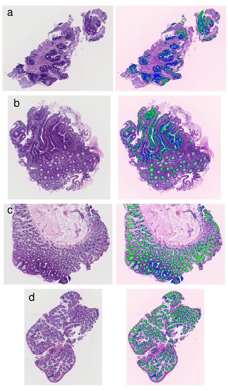
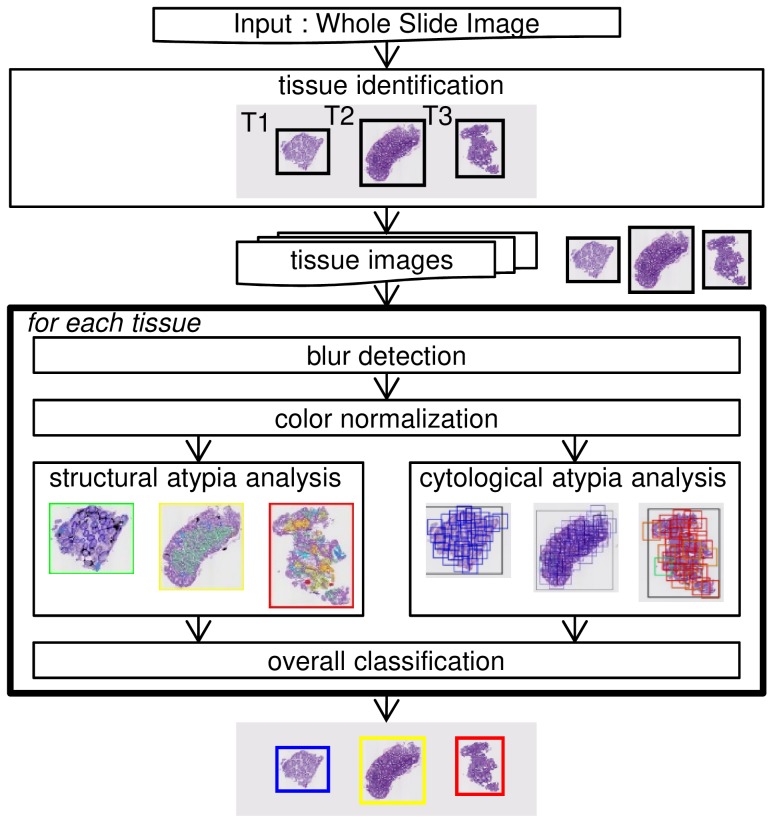
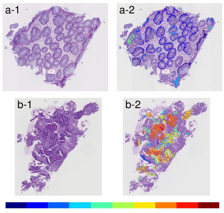
An automated image analysis system, e-Pathologist, was developed to improve the quality of colorectal biopsy diagnostics in routine pathology practice.

OBJECTIVE

The aim of the study was to evaluate the classification accuracy of the e-Pathologist image analysis software in the setting of routine pathology practice in two institutions.

MATERIALS AND METHODS

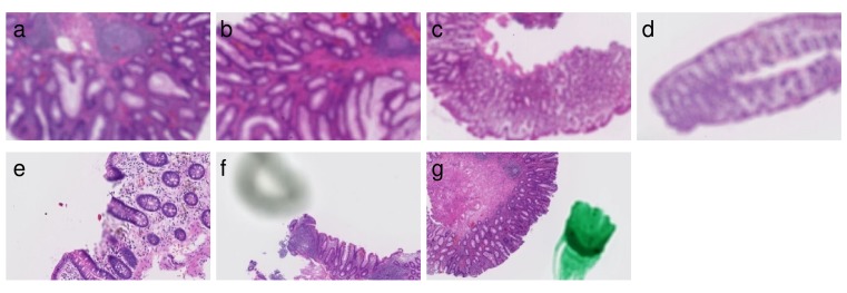
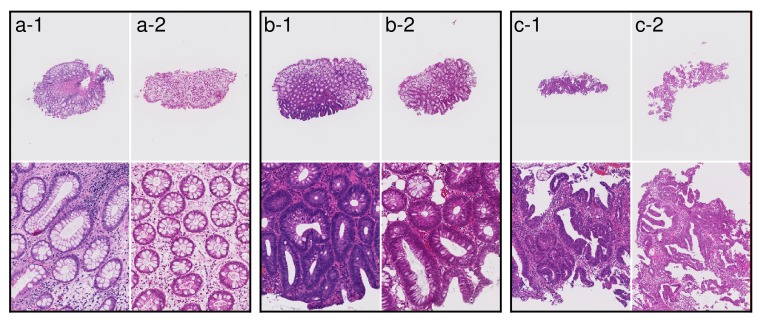
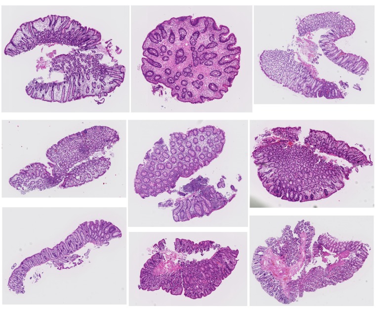
In total, 1328 colorectal tissue specimens were consecutively obtained from two hospitals (1077 tissues from Tokyo hospital, and 251 tissues from East hospital) and the stained specimen slides were anonymized and digitized. At least two experienced gastrointestinal pathologists evaluated each slide for pathological diagnosis. We compared the 3-tier classification results (carcinoma or suspicion of carcinoma, adenoma, and lastly negative for a neoplastic lesion) between the human pathologists and that of e-Pathologist.

RESULTS

For the Tokyo hospital specimens, all carcinoma tissues were correctly classified (n=112), and 9.9% (80/810) of the adenoma tissues were incorrectly classified as negative. For the East hospital specimens, 0 out of the 51 adenoma tissues were incorrectly classified as negative while 9.3% (11/118) of the carcinoma tissues were incorrectly classified as either adenoma, or negative. For the Tokyo and East hospital datasets, the undetected rate of carcinoma, undetected rate of adenoma, and over-detected proportion were 0% and 9.3%, 9.9% and 0%, and 36.1% and 27.1%, respectively.

CONCLUSIONS

This image analysis system requires some improvements; however, it has the potential to assist pathologists in quality improvement of routine pathological practice in the not too distant future.

摘要

背景

开发了一种自动化图像分析系统e-Pathologist,以提高常规病理实践中结直肠活检诊断的质量。

目的

本研究的目的是评估e-Pathologist图像分析软件在两家机构常规病理实践中的分类准确性。

材料和方法

总共从两家医院连续获取了1328份结直肠组织标本(东京医院1077份组织,东方医院251份组织),对染色的标本玻片进行匿名化处理并数字化。至少两名经验丰富的胃肠病理学家对每张玻片进行病理诊断评估。我们比较了人类病理学家和e-Pathologist的三级分类结果(癌或疑似癌、腺瘤,最后为肿瘤性病变阴性)。

结果

对于东京医院的标本,所有癌组织均被正确分类(n = 112),9.9%(80/810)的腺瘤组织被错误分类为阴性。对于东方医院的标本,51份腺瘤组织中没有一份被错误分类为阴性,而9.3%(11/118)的癌组织被错误分类为腺瘤或阴性。对于东京医院和东方医院的数据集,癌的未检出率、腺瘤的未检出率和过度检出比例分别为0%和9.3%、9.9%和0%、36.1%和27.1%。

结论

该图像分析系统需要一些改进;然而,在不久的将来,它有可能帮助病理学家提高常规病理实践的质量。

https://cdn.ncbi.nlm.nih.gov/pmc/blobs/a2ba/5710880/af2bb4801c5b/oncotarget-08-90719-g007.jpg
https://cdn.ncbi.nlm.nih.gov/pmc/blobs/a2ba/5710880/e68cc3223fd5/oncotarget-08-90719-g001.jpg
https://cdn.ncbi.nlm.nih.gov/pmc/blobs/a2ba/5710880/24c36d56bacd/oncotarget-08-90719-g002.jpg
https://cdn.ncbi.nlm.nih.gov/pmc/blobs/a2ba/5710880/0965c4700e29/oncotarget-08-90719-g003.jpg
https://cdn.ncbi.nlm.nih.gov/pmc/blobs/a2ba/5710880/ef46e9326e76/oncotarget-08-90719-g004.jpg
https://cdn.ncbi.nlm.nih.gov/pmc/blobs/a2ba/5710880/b77bd29a2686/oncotarget-08-90719-g005.jpg
https://cdn.ncbi.nlm.nih.gov/pmc/blobs/a2ba/5710880/f89bf881cc20/oncotarget-08-90719-g006.jpg
https://cdn.ncbi.nlm.nih.gov/pmc/blobs/a2ba/5710880/af2bb4801c5b/oncotarget-08-90719-g007.jpg

相似文献

1
Automated histological classification of whole slide images of colorectal biopsy specimens.结直肠活检标本全切片图像的自动化组织学分类
Oncotarget. 2017 Oct 12;8(53):90719-90729. doi: 10.18632/oncotarget.21819. eCollection 2017 Oct 31.
2
Automated histological classification of whole-slide images of gastric biopsy specimens.胃活检标本全切片图像的自动化组织学分类。
Gastric Cancer. 2018 Mar;21(2):249-257. doi: 10.1007/s10120-017-0731-8. Epub 2017 Jun 2.
3
Evaluation of a Deep Neural Network for Automated Classification of Colorectal Polyps on Histopathologic Slides.基于病理切片的深度学习神经网络自动结直肠息肉分类评估。
JAMA Netw Open. 2020 Apr 1;3(4):e203398. doi: 10.1001/jamanetworkopen.2020.3398.
4
MIST: multiple instance learning network based on Swin Transformer for whole slide image classification of colorectal adenomas.MIST:基于Swin Transformer的多实例学习网络用于结直肠腺瘤全切片图像分类
J Pathol. 2023 Feb;259(2):125-135. doi: 10.1002/path.6027. Epub 2022 Dec 7.
5
Improving quality control in the routine practice for histopathological interpretation of gastrointestinal endoscopic biopsies using artificial intelligence.利用人工智能提高胃肠道内镜活检组织病理解释常规实践中的质量控制。
PLoS One. 2022 Dec 15;17(12):e0278542. doi: 10.1371/journal.pone.0278542. eCollection 2022.
6
Assessment of Machine Learning of Breast Pathology Structures for Automated Differentiation of Breast Cancer and High-Risk Proliferative Lesions.基于机器学习的乳腺病理学结构评估在乳腺癌和高危增殖性病变自动鉴别中的应用。
JAMA Netw Open. 2019 Aug 2;2(8):e198777. doi: 10.1001/jamanetworkopen.2019.8777.
7
iPathology cockpit diagnostic station: validation according to College of American Pathologists Pathology and Laboratory Quality Center recommendation at the Hospital Trust and University of Verona.iPathology 座舱诊断工作站:根据美国病理学家学会病理与实验室质量中心的建议,在维罗纳医院信托和大学进行验证。
Diagn Pathol. 2014;9 Suppl 1(Suppl 1):S12. doi: 10.1186/1746-1596-9-S1-S12. Epub 2014 Dec 19.
8
AI (artificial intelligence) in histopathology--from image analysis to automated diagnosis.组织病理学中的人工智能——从图像分析到自动诊断。
Folia Histochem Cytobiol. 2009 Jan;47(3):355-61. doi: 10.2478/v10042-009-0087-y.
9
International comparability of the pathological diagnosis for early cancer of the digestive tract: Munich meeting.消化道早期癌病理诊断的国际可比性:慕尼黑会议
J Gastroenterol. 2000;35 Suppl 12:102-10.
10
Computer-aided evaluation of neuroblastoma on whole-slide histology images: Classifying grade of neuroblastic differentiation.基于全切片组织学图像的神经母细胞瘤计算机辅助评估:神经母细胞分化程度分类
Pattern Recognit. 2009 Jun;42(6):1080-1092. doi: 10.1016/j.patcog.2008.10.035.

引用本文的文献

1
Automated recognition of glomerular lesions in the kidneys of mice by using deep learning.利用深度学习自动识别小鼠肾脏中的肾小球病变。
J Pathol Inform. 2022 Jul 28;13:100129. doi: 10.1016/j.jpi.2022.100129. eCollection 2022.
2
The seen and the unseen: Molecular classification and image based-analysis of gastrointestinal cancers.可见与不可见:胃肠道癌症的分子分类及基于图像的分析
Comput Struct Biotechnol J. 2022 Sep 12;20:5065-5075. doi: 10.1016/j.csbj.2022.09.010. eCollection 2022.
3
Automated histological classification for digital pathology images of colonoscopy specimen via deep learning.

本文引用的文献

1
Japanese Society for Cancer of the Colon and Rectum (JSCCR) Guidelines 2014 for treatment of colorectal cancer.日本结直肠癌学会(JSCCR)2014年结直肠癌治疗指南。
Int J Clin Oncol. 2015 Apr;20(2):207-39. doi: 10.1007/s10147-015-0801-z. Epub 2015 Mar 18.
2
A hybrid classification model for digital pathology using structural and statistical pattern recognition.基于结构和统计模式识别的数字病理学混合分类模型。
IEEE Trans Med Imaging. 2013 Feb;32(2):474-83. doi: 10.1109/TMI.2012.2230186. Epub 2012 Nov 27.
3
Color graphs for automated cancer diagnosis and grading.
基于深度学习的结肠镜标本数字病理学图像的自动化组织学分类。
Sci Rep. 2022 Jul 27;12(1):12804. doi: 10.1038/s41598-022-16885-x.
4
Paper-based genetic assays with bioconjugated gold nanorods and an automated readout pipeline.基于金纳米棒的生物共轭物和自动化读取的纸质基因检测。
Sci Rep. 2022 Apr 13;12(1):6223. doi: 10.1038/s41598-022-10227-7.
5
Development of artificial intelligence technology in diagnosis, treatment, and prognosis of colorectal cancer.人工智能技术在结直肠癌诊断、治疗及预后方面的发展
World J Gastrointest Oncol. 2022 Jan 15;14(1):124-152. doi: 10.4251/wjgo.v14.i1.124.
6
Deep learning-based histopathological segmentation for whole slide images of colorectal cancer in a compressed domain.基于深度学习的结直肠癌全切片图像在压缩域中的病理分割。
Sci Rep. 2021 Nov 18;11(1):22520. doi: 10.1038/s41598-021-01905-z.
7
CAD systems for colorectal cancer from WSI are still not ready for clinical acceptance.基于 WSI 的结直肠癌 CAD 系统尚未准备好临床应用。
Sci Rep. 2021 Jul 13;11(1):14358. doi: 10.1038/s41598-021-93746-z.
8
Requirements for implementation of artificial intelligence in the practice of gastrointestinal pathology.人工智能在胃肠病学实践中的应用要求。
World J Gastroenterol. 2021 Jun 7;27(21):2818-2833. doi: 10.3748/wjg.v27.i21.2818.
9
Current Trends of Artificial Intelligence for Colorectal Cancer Pathology Image Analysis: A Systematic Review.人工智能用于结直肠癌病理图像分析的当前趋势:一项系统综述
Cancers (Basel). 2020 Jul 13;12(7):1884. doi: 10.3390/cancers12071884.
10
A review of the application of deep learning in medical image classification and segmentation.深度学习在医学图像分类与分割中的应用综述。
Ann Transl Med. 2020 Jun;8(11):713. doi: 10.21037/atm.2020.02.44.
用于癌症自动诊断和分级的彩色图谱。
IEEE Trans Biomed Eng. 2010 Mar;57(3):665-74. doi: 10.1109/TBME.2009.2033804. Epub 2009 Oct 20.
4
The Vienna classification of gastrointestinal epithelial neoplasia.胃肠道上皮性肿瘤的维也纳分类
Gut. 2000 Aug;47(2):251-5. doi: 10.1136/gut.47.2.251.