Eglinger Jan, Karsjens Haiko, Lammert Eckhard
Institute of Metabolic Physiology, Heinrich-Heine University, Düsseldorf, Germany.
Institute for Beta Cell Biology, Leibniz Center for Diabetes Research, German Diabetes Center (DDZ), Düsseldorf, Germany.
Inflamm Regen. 2017 Jan 18;37:2. doi: 10.1186/s41232-016-0033-2. eCollection 2017.
Pericytes, surrounding the endothelium, fulfill diverse functions that are crucial for vascular homeostasis. The loss of pericytes is associated with pathologies, such as diabetic retinopathy and Alzheimer's disease. Thus, there exists a need for an experimental system that combines pharmacologic manipulation and quantification of pericyte coverage during sprouting angiogenesis. Here, we describe an in vitro angiogenesis assay that develops lumenized vascular sprouts composed of endothelial cells enveloped by pericytes, with the additional ability to comparatively screen the effect of multiple small molecules simultaneously. For automated analysis, we also present an ImageJ plugin tool we developed to quantify sprout morphology and pericyte coverage.
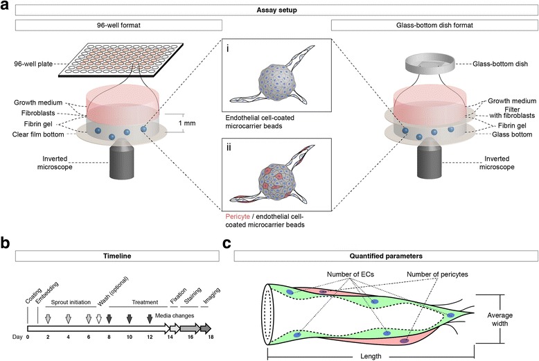
Human umbilical vein endothelial cells and human brain vascular pericytes were coated on microcarrier beads and embedded in fibrin gels in a 96-well plate to form lumenized vascular sprouts. After treatment with pharmacologic compounds, sprouts were fixed, stained, and imaged via optical z-sections over the area of each well. The maximum intensity projections of these images were stitched together to form montages of the wells, and those montages were processed and analyzed.
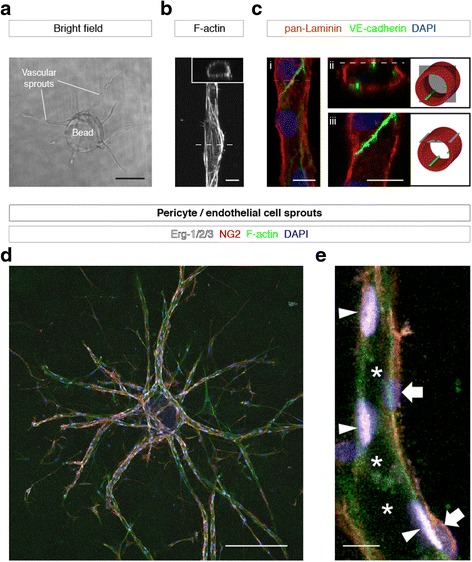
Vascular sprouts formed within 4-12 days and contained a patent lumen surrounded by a layer of human endothelial cells and pericytes. Using our workflow and image analysis, pericyte coverage after treatment with various compounds was successfully quantified.
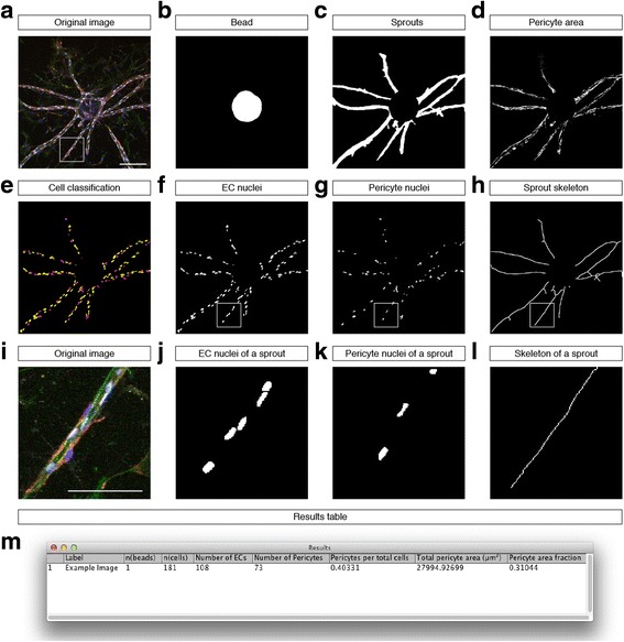
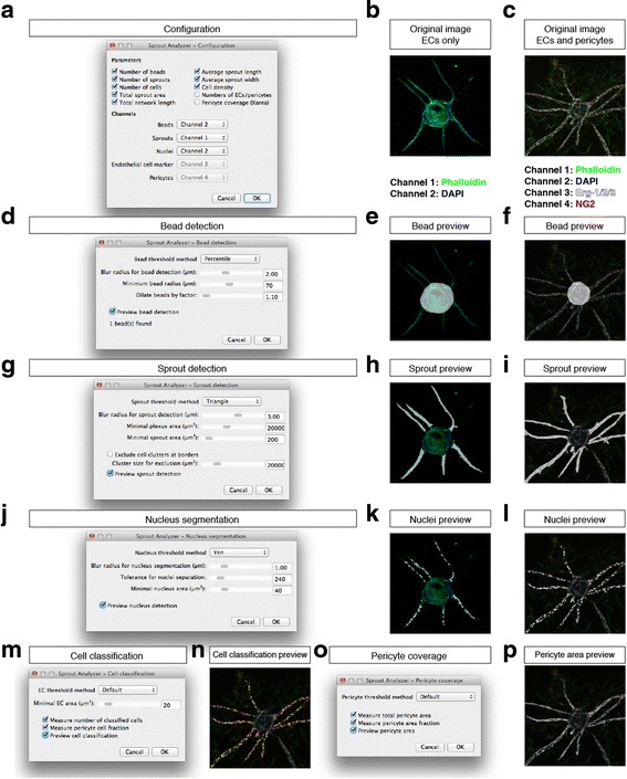
Here we present a robust in vitro assay using primary human vascular cells that allows researchers to analyze the effects of multiple compounds on sprouting angiogenesis and pericyte coverage. Our ImageJ plugin offers automated evaluation across multiple different vascular parameters, such as sprout length, cell density, branch points, and pericyte coverage.
周细胞围绕在内皮细胞周围,发挥着对血管稳态至关重要的多种功能。周细胞的丧失与诸如糖尿病视网膜病变和阿尔茨海默病等病理状况相关。因此,需要一种实验系统,该系统能够在发芽血管生成过程中结合药理学操作和周细胞覆盖率的量化。在此,我们描述了一种体外血管生成测定法,该方法可形成由周细胞包裹的内皮细胞组成的有腔血管芽,并且还具有同时比较筛选多种小分子作用效果的能力。为了进行自动化分析,我们还展示了我们开发的一种ImageJ插件工具,用于量化芽的形态和周细胞覆盖率。
将人脐静脉内皮细胞和人脑血管周细胞包被在微载体珠上,并嵌入96孔板中的纤维蛋白凝胶中以形成有腔血管芽。在用药物化合物处理后,将芽固定、染色,并通过对每个孔区域的光学z轴切片进行成像。这些图像的最大强度投影拼接在一起形成孔的蒙太奇图像,然后对这些蒙太奇图像进行处理和分析。
血管芽在4 - 12天内形成,并包含一个由一层人内皮细胞和周细胞包围的开放管腔。使用我们的工作流程和图像分析,成功量化了用各种化合物处理后的周细胞覆盖率。
在此我们展示了一种使用原代人血管细胞的强大体外测定法,该方法使研究人员能够分析多种化合物对发芽血管生成和周细胞覆盖率的影响。我们的ImageJ插件可对多个不同的血管参数进行自动评估,例如芽长度、细胞密度、分支点和周细胞覆盖率。