Department of Rheumatology & Clinical Immunology, Amsterdam Rheumatology & Immunology Center, Amsterdam University Medical Centers, University of Amsterdam, Amsterdam, Netherlands.
Department of Experimental Immunology, Amsterdam University Medical Centers, University of Amsterdam, Amsterdam, Netherlands.
Front Immunol. 2023 Jul 20;14:1188835. doi: 10.3389/fimmu.2023.1188835. eCollection 2023.
Rheumatoid Arthritis (RA) is a progressive and systemic autoimmune disorder associated with chronic and destructive joint inflammation. The hallmarks of joint synovial inflammation are cellular proliferation, extensive neoangiogenesis and infiltration of immune cells, including macrophages. approaches simulating RA synovial tissue are crucial in preclinical and translational research to evaluate novel diagnostic and/or therapeutic markers. Two-dimensional (2D) settings present very limited physiological proximity as they cannot recapitulate cell-cell and cell-matrix interactions occurring in the three-dimensional (3D) tissue compartment. Here, we present the engineering of a spheroid-based model of RA synovial tissue which mimics 3D interactions between cells and pro-inflammatory mediators present in the inflamed synovium.
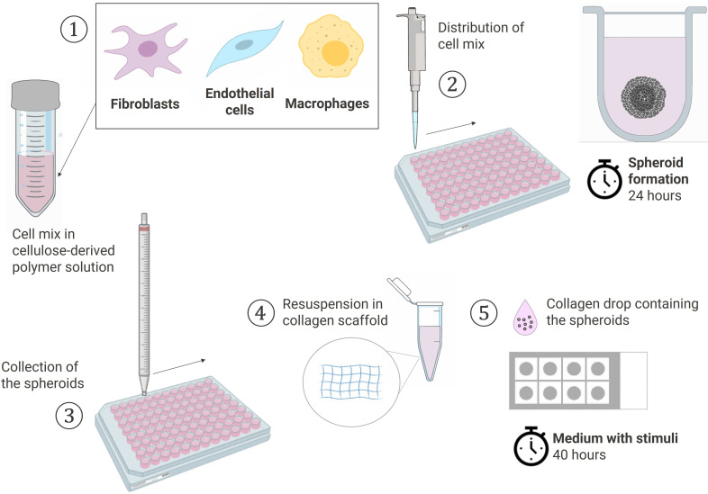
Spheroids were generated by culturing RA fibroblast-like-synoviocytes (RAFLS), human umbilical vein endothelial cells (ECs) and monocyte-derived macrophages in a collagen-based 3D scaffold. The spheroids were cultured in the presence or absence of vascular endothelial growth factor (VEGF) and fibroblast growth factor 2 (bFGF) or RA synovial fluid (SF). Spheroid expansion and cell migration were quantified for all conditions using confocal microscopy and digital image analysis.
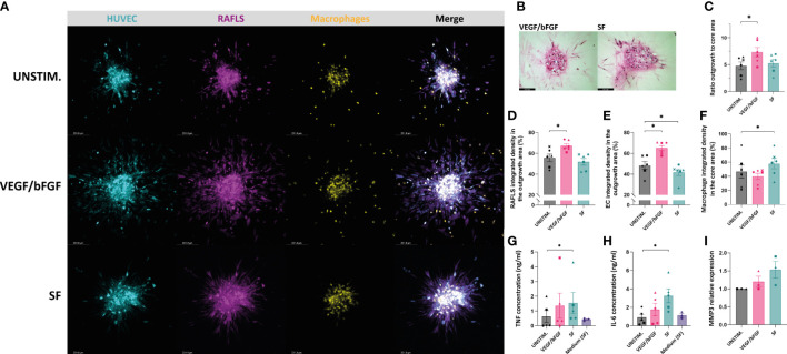
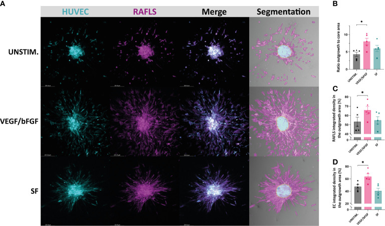
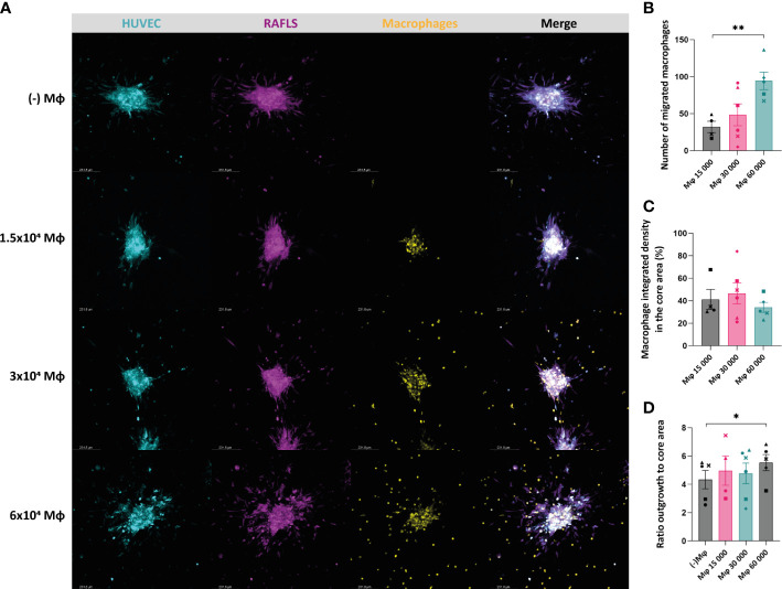
A novel approach using machine learning was developed to quantify spheroid outgrowth and used to reexamine the existing spheroid-based model of RA synovial angiogenesis consisting of ECs and RAFLS. A 2-fold increase in the spheroid outgrowth ratio was demonstrated upon VEGF/bFGF stimulation (<0.05). The addition of macrophages within the spheroid structure (3.75x10 RAFLS, 7.5x10 ECs and 3.0x10 macrophages) resulted in good incorporation of the new cell type. The addition of VEGF/bFGF significantly induced spheroid outgrowth (<0.05) in the new system. SF stimulation enhanced containment of macrophages within the spheroids.
We present a novel spheroid based model consisting of RAFLS, ECs and macrophages that reflects the RA synovial tissue microenvironment. This model may be used to dissect the role of specific cell types in inflammatory responses in RA, to study specific signaling pathways involved in the disease pathogenesis and examine the effects of novel diagnostic (molecular imaging) and therapeutic compounds, including small molecule inhibitors and biologics.
类风湿关节炎(RA)是一种与慢性、破坏性关节炎症相关的进行性、系统性自身免疫性疾病。关节滑膜炎症的标志是细胞增殖、广泛的新血管生成和免疫细胞浸润,包括巨噬细胞。模拟 RA 滑膜组织的方法在临床前和转化研究中至关重要,可用于评估新的诊断和/或治疗标志物。二维(2D)设置由于不能再现发生在三维(3D)组织隔室中的细胞-细胞和细胞-基质相互作用,因此提供的生理相似性非常有限。在这里,我们提出了一种基于球体的 RA 滑膜组织模型的工程设计,该模型模拟了细胞之间以及炎症滑膜中存在的促炎介质之间的 3D 相互作用。
通过在胶原基 3D 支架中培养 RA 成纤维样滑膜细胞(RAFLS)、人脐静脉内皮细胞(ECs)和单核细胞衍生的巨噬细胞来生成球体。在存在或不存在血管内皮生长因子(VEGF)和碱性成纤维细胞生长因子 2(bFGF)或 RA 滑膜液(SF)的情况下培养球体。使用共聚焦显微镜和数字图像分析定量所有条件下球体的扩展和细胞迁移。
开发了一种使用机器学习的新方法来量化球体的生长,并用于重新检查由 ECs 和 RAFLS 组成的现有的 RA 滑膜血管生成球体模型。在 VEGF/bFGF 刺激下,球体生长率增加了 2 倍(<0.05)。在球体结构中添加巨噬细胞(3.75x10 RAFLS、7.5x10 ECs 和 3.0x10 巨噬细胞)可很好地掺入新细胞类型。在新系统中,添加 VEGF/bFGF 可显著诱导球体生长(<0.05)。SF 刺激增强了巨噬细胞在球体中的包封。
我们提出了一种新的基于球体的模型,由 RAFLS、ECs 和巨噬细胞组成,反映了 RA 滑膜组织的微环境。该模型可用于剖析特定细胞类型在 RA 炎症反应中的作用,研究疾病发病机制中涉及的特定信号通路,并研究新型诊断(分子成像)和治疗化合物的作用,包括小分子抑制剂和生物制剂。