Kang Kai B, Lawrence Brian D, Gao X Raymond, Luo Yuncin, Zhou Qiang, Liu Aihong, Guaiquil Victor H, Rosenblatt Mark I
Department of Ophthalmology and Visual Sciences, Illinois Eye and Ear Infirmary, University of Illinois at Chicago, Chicago, Illinois, United States.
Department of Ophthalmology, Weill Cornell Medical College, New York, New York, United States.
Invest Ophthalmol Vis Sci. 2017 Dec 1;58(14):6388-6398. doi: 10.1167/iovs.17-22213.
Corneal basement membrane has topographical features that provide biophysical cues to direct cell adherence, migration, and proliferation. In this study, we hypothesize that varying topographic pitch created on silk films can alter epithelial cell morphology, adhesion, and the genetic expression involved in cytoskeletal dynamics-related pathways.
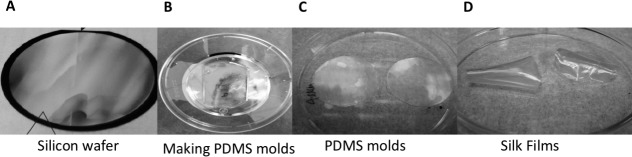
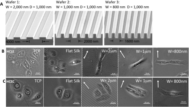
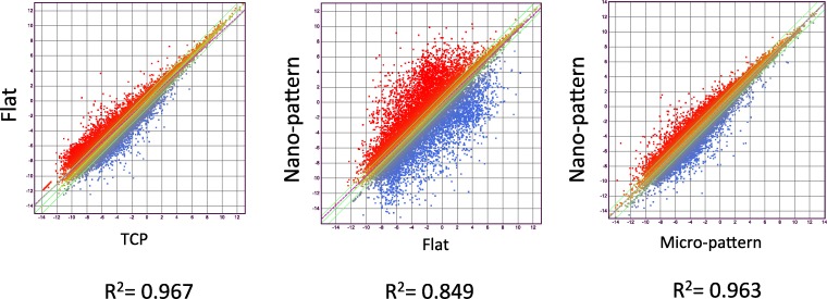
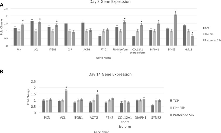
Silicon wafers with parallel ridge widths of 2000, 1000, and 800 nm were produced and used to pattern silk films via soft lithography. Human corneal epithelial cells were cultured onto silk. After 72 hours of incubation, images were taken to study cell morphology and alignment. Cytoskeletal structures were studied by immunofluorescent staining. RNA was collected from cultured cells to perform RNA-Seq transcriptome analysis using the Illumina Hiseq 2500 sequencing system. Differentially expressed genes were identified using DNAstar Qseq then verified using quantitative real-time PCR. These genes were used to perform pathway analyses using Ingenuity Pathways Analysis.
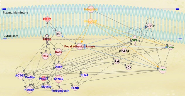
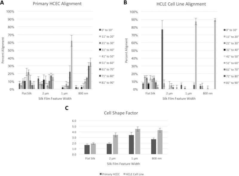
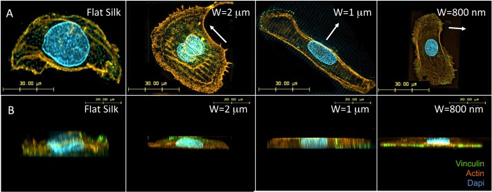
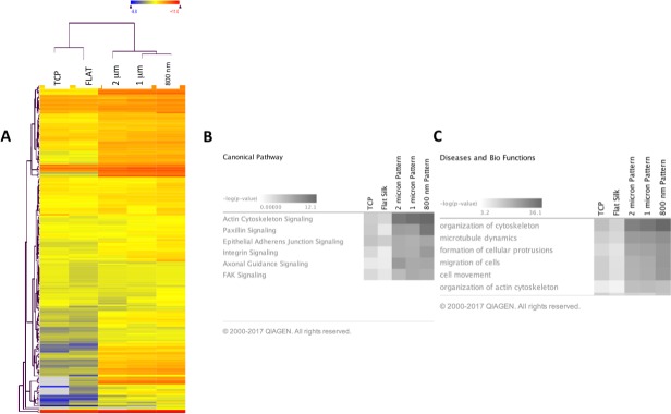
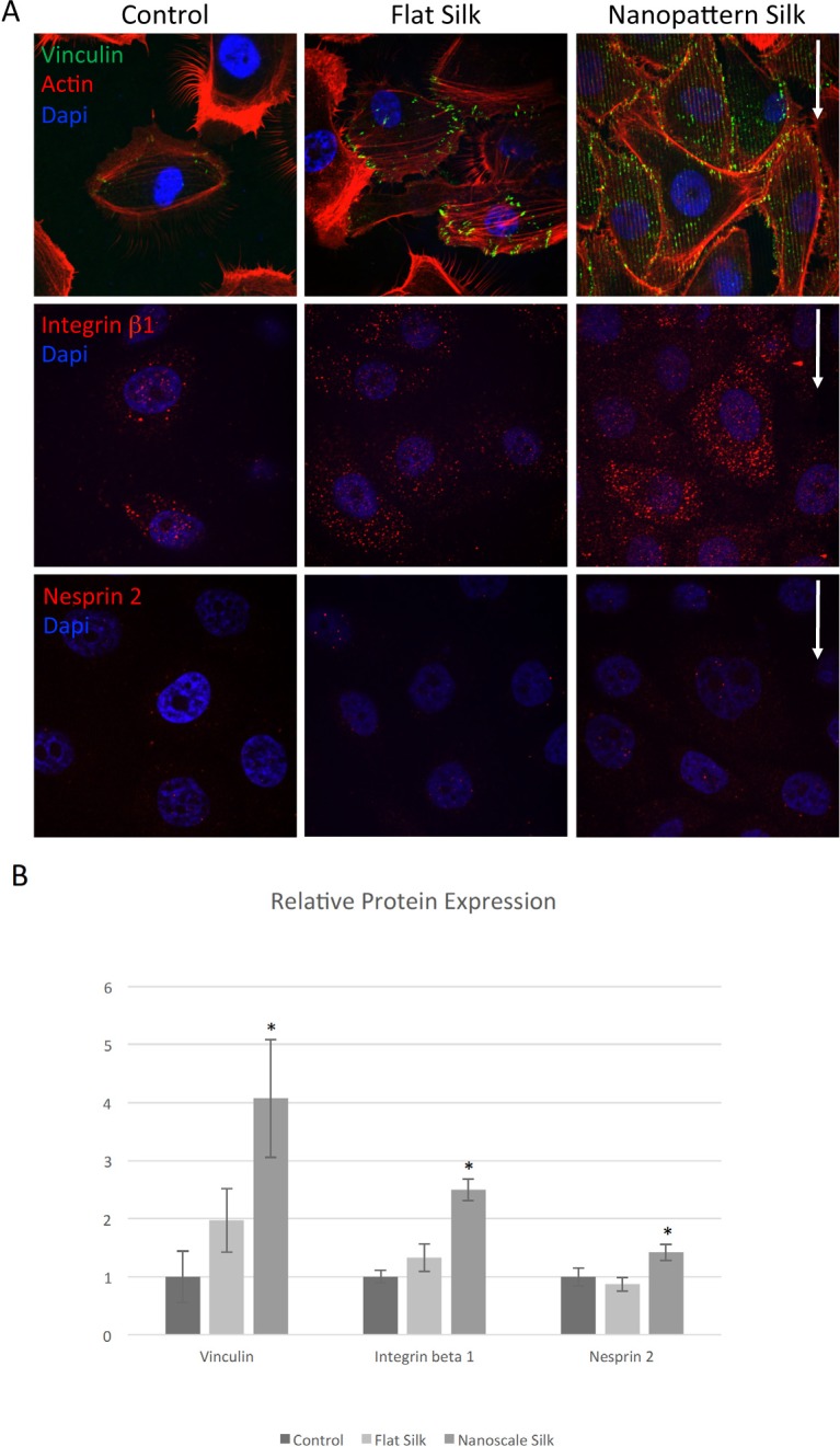
Primary human corneal epithelial cell alignment to the surface pattern was the greatest on 1000-nm features. Fluorescent microscopy of f-actin staining showed cell cytoskeleton alignment either in parallel (2000 nm) or perpendicular (1000 and 800 nm) to the long feature axis. Z-stack projection of vinculin staining indicated increased focal adhesion formation localized on the cellular basal surface. RNA-seq analysis revealed differentially expressed genes involved in actin organization, integrin signaling, and focal adhesion kinase signaling (-log (P)>5).
Patterned silk film substrates may serve as a scaffold and provide biophysical cues to corneal epithelial cells that change their gene expression, alter cellular adherence, morphology, and may offer a promising customizable material for use in ocular surface repair.
角膜基底膜具有地形学特征,可提供生物物理线索以指导细胞黏附、迁移和增殖。在本研究中,我们假设在丝膜上创建的不同地形间距可改变上皮细胞形态、黏附以及细胞骨架动力学相关途径中涉及的基因表达。
制作平行脊宽为2000、1000和800 nm的硅片,并通过软光刻技术用于对丝膜进行图案化。将人角膜上皮细胞培养在丝膜上。孵育72小时后,拍摄图像以研究细胞形态和排列。通过免疫荧光染色研究细胞骨架结构。从培养的细胞中收集RNA,使用Illumina Hiseq 2500测序系统进行RNA测序转录组分析。使用DNAstar Qseq鉴定差异表达基因,然后使用定量实时PCR进行验证。这些基因用于使用Ingenuity Pathways Analysis进行通路分析。
原代人角膜上皮细胞在1000 nm特征上与表面图案的排列最为明显。f-肌动蛋白染色的荧光显微镜显示细胞细胞骨架与长特征轴平行(2000 nm)或垂直(1000和800 nm)排列。纽蛋白染色的Z轴堆叠投影表明细胞基底表面局部形成的粘着斑增加。RNA测序分析揭示了参与肌动蛋白组织、整合素信号传导和粘着斑激酶信号传导的差异表达基因(-log(P)>5)。
图案化的丝膜基质可作为支架,并为角膜上皮细胞提供生物物理线索,从而改变其基因表达、改变细胞黏附、形态,并可能为眼表修复提供一种有前景的可定制材料。