Department of Ophthalmology and Visual Sciences, Illinois Eye and Ear Infirmary, University of Illinois at Chicago, Chicago, IL, USA.
Department of Ophthalmology, Weill Cornell Medical College, New York, NY, USA.
Sci Rep. 2019 Feb 6;9(1):1507. doi: 10.1038/s41598-018-37804-z.
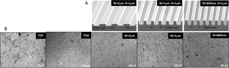
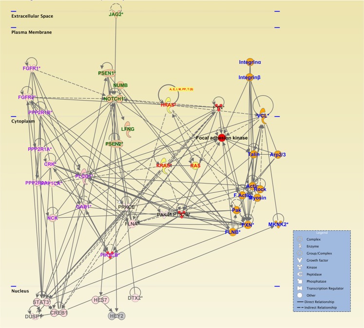
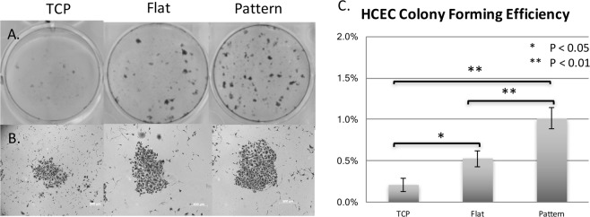
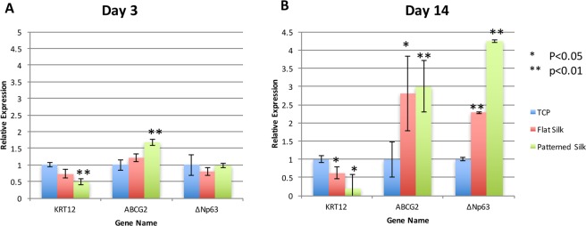
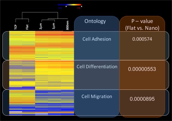
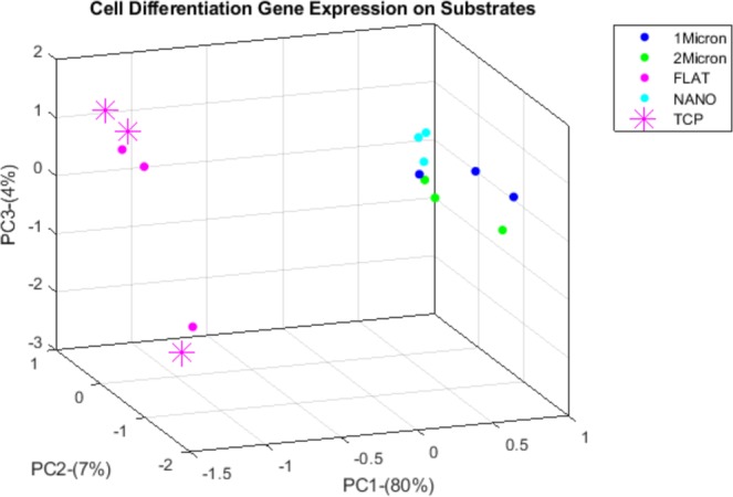
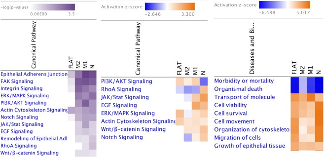
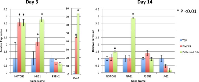
We previously reported that micro- and nano-scale topographic pitch created on silk films mimic features of the corneal basement membrane by providing biophysical cues to direct corneal epithelial cell adherence and migration. However, the effect of these topographical features on corneal limbal epithelial cell differentiation has not been explored. We hypothesize in the current study that various topographical pitch created on silk may affect corneal epithelial stem cell differentiation and alter the expression of genes involved in cell differentiation and self-renewal. We patterned silk films with different topographic pitch via soft lithography and observed human corneal limbal epithelial cell behavior. Colony forming assay demonstrated increased colony forming efficiency on patterned silk films. Cells cultured on nanoscale patterned silk films also expressed lower levels of putative keratocyte differentiation markers and higher levels of putative limbal stem cell markers. RNA-Seq analysis further implicated the involvement of pathways related to stem cell differentiation and self-renewal, including Notch, ERK/MAPK and Wnt/β-catenin signaling. We conclude that patterned silk film substrates can be used as scaffolds and provide biophysical cues to corneal limbal stem cells that may maintain corneal epithelial stem cells at a less differentiated state.
我们之前曾报道过,在丝膜上形成的微观和纳米级的形貌峰谷结构模仿了角膜基底层的特征,为角膜上皮细胞的黏附和迁移提供了生物物理线索。然而,这些形貌特征对角膜缘上皮细胞分化的影响尚未得到探索。在本研究中,我们假设在丝上形成的不同形貌峰谷可能会影响角膜上皮干细胞的分化,并改变与细胞分化和自我更新相关的基因的表达。我们通过软光刻技术在丝膜上形成具有不同形貌峰谷的图案,并观察人角膜缘上皮细胞的行为。集落形成实验表明,在图案化丝膜上的集落形成效率增加。在纳米级图案化丝膜上培养的细胞也表达了较低水平的推测角膜细胞分化标志物和较高水平的推测角膜缘干细胞标志物。RNA-Seq 分析进一步表明,涉及干细胞分化和自我更新的途径,包括 Notch、ERK/MAPK 和 Wnt/β-catenin 信号通路的参与。我们得出结论,图案化丝膜底物可用作支架,并为角膜缘干细胞提供生物物理线索,这可能使角膜上皮干细胞保持在较低分化状态。