Computational Biology Department, School of Computer Science, Carnegie Mellon University, Pittsburgh 15213, USA.
Division of Structural Biology, Wellcome Trust Centre for Human Genetics, University of Oxford, Oxford OX3 7BN, UK; Cryo-electron Microscopy, Bijvoet Center for Biomolecular Research, Utrecht University, Utrecht, Netherlands.
J Struct Biol. 2018 May;202(2):150-160. doi: 10.1016/j.jsb.2017.12.015. Epub 2017 Dec 28.
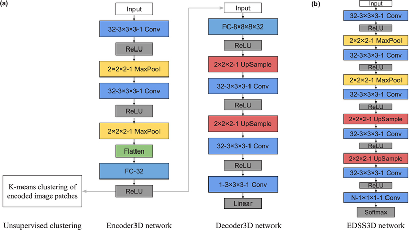
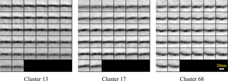
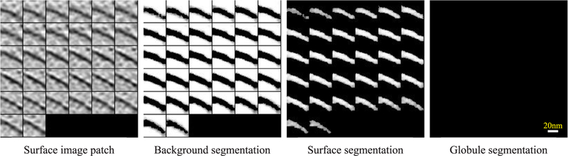
Cellular electron cryo-tomography enables the 3D visualization of cellular organization in the near-native state and at submolecular resolution. However, the contents of cellular tomograms are often complex, making it difficult to automatically isolate different in situ cellular components. In this paper, we propose a convolutional autoencoder-based unsupervised approach to provide a coarse grouping of 3D small subvolumes extracted from tomograms. We demonstrate that the autoencoder can be used for efficient and coarse characterization of features of macromolecular complexes and surfaces, such as membranes. In addition, the autoencoder can be used to detect non-cellular features related to sample preparation and data collection, such as carbon edges from the grid and tomogram boundaries. The autoencoder is also able to detect patterns that may indicate spatial interactions between cellular components. Furthermore, we demonstrate that our autoencoder can be used for weakly supervised semantic segmentation of cellular components, requiring a very small amount of manual annotation.
细胞电子断层扫描技术能够以近天然状态和亚分子分辨率对细胞组织进行 3D 可视化。然而,细胞断层图像的内容通常较为复杂,因此很难自动分离不同的原位细胞成分。在本文中,我们提出了一种基于卷积自动编码器的无监督方法,用于对从断层图像中提取的 3D 小体积进行粗略分组。我们证明了自动编码器可用于高效且粗略地描述大分子复合物和表面(如膜)的特征。此外,自动编码器还可用于检测与样品制备和数据采集相关的非细胞特征,例如网格的碳边缘和断层图像的边界。自动编码器还能够检测可能表明细胞成分之间空间相互作用的模式。此外,我们证明了我们的自动编码器可用于细胞成分的弱监督语义分割,仅需要少量的手动注释。