• 文献检索
  • 文档翻译
  • 深度研究
  • 学术资讯
  • Suppr Zotero 插件Zotero 插件
  • 邀请有礼
  • 套餐&价格
  • 历史记录
应用&插件
Suppr Zotero 插件Zotero 插件浏览器插件Mac 客户端Windows 客户端微信小程序
定价
高级版会员购买积分包购买API积分包
服务
文献检索文档翻译深度研究API 文档MCP 服务
关于我们
关于 Suppr公司介绍联系我们用户协议隐私条款
关注我们

Suppr 超能文献

核心技术专利:CN118964589B侵权必究
粤ICP备2023148730 号-1Suppr @ 2026

文献检索

告别复杂PubMed语法,用中文像聊天一样搜索,搜遍4000万医学文献。AI智能推荐,让科研检索更轻松。

立即免费搜索

文件翻译

保留排版,准确专业,支持PDF/Word/PPT等文件格式,支持 12+语言互译。

免费翻译文档

深度研究

AI帮你快速写综述,25分钟生成高质量综述,智能提取关键信息,辅助科研写作。

立即免费体验

通过冷冻电镜断层扫描和相关显微镜技术对兴奋性和抑制性突触的分化和特征进行研究。

Differentiation and Characterization of Excitatory and Inhibitory Synapses by Cryo-electron Tomography and Correlative Microscopy.

机构信息

National Laboratory for Physical Sciences at the Microscale.

School of Life Sciences.

出版信息

J Neurosci. 2018 Feb 7;38(6):1493-1510. doi: 10.1523/JNEUROSCI.1548-17.2017. Epub 2018 Jan 8.

DOI:10.1523/JNEUROSCI.1548-17.2017
PMID:29311144
原文链接:https://pmc.ncbi.nlm.nih.gov/articles/PMC5815350/
Abstract

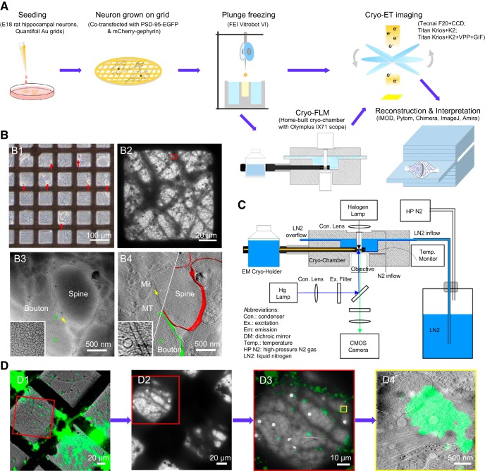
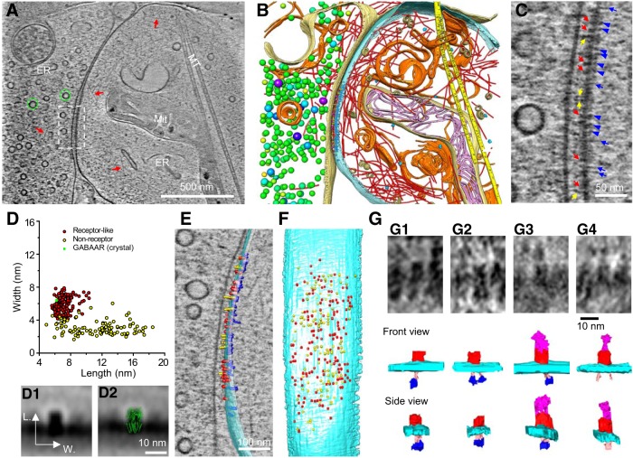
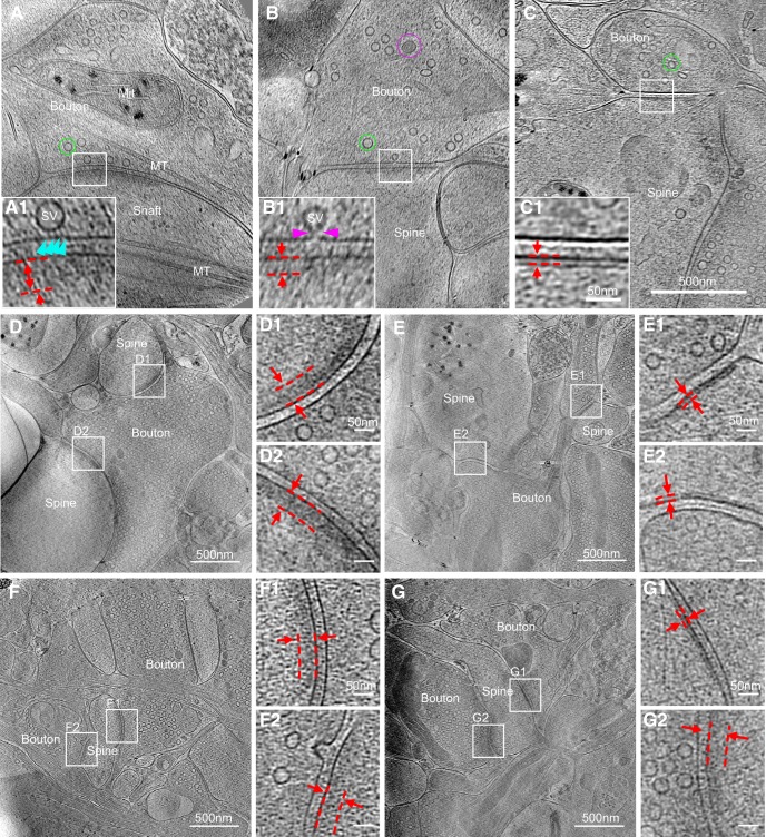
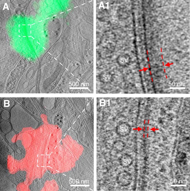
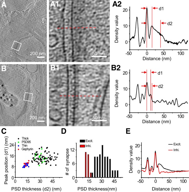
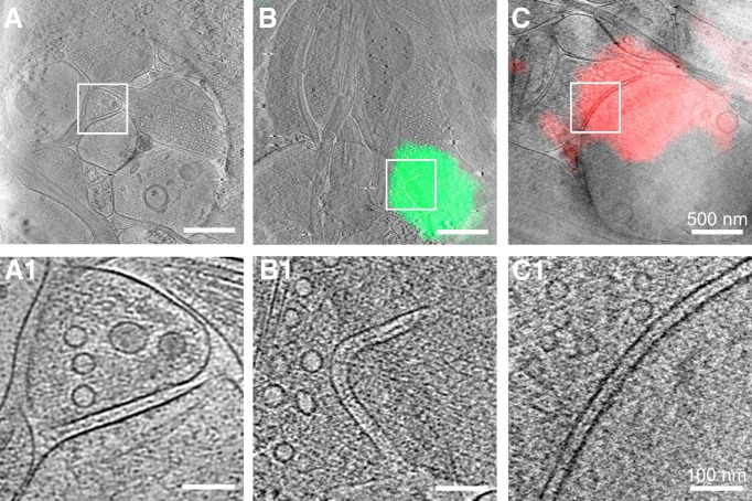
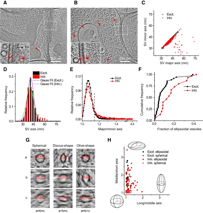
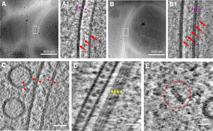
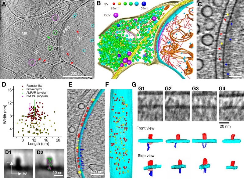
As key functional units in neural circuits, different types of neuronal synapses play distinct roles in brain information processing, learning, and memory. Synaptic abnormalities are believed to underlie various neurological and psychiatric disorders. Here, by combining cryo-electron tomography and cryo-correlative light and electron microscopy, we distinguished intact excitatory and inhibitory synapses of cultured hippocampal neurons, and visualized the 3D organization of synaptic organelles and macromolecules in their native state. Quantitative analyses of >100 synaptic tomograms reveal that excitatory synapses contain a mesh-like postsynaptic density (PSD) with thickness ranging from 20 to 50 nm. In contrast, the PSD in inhibitory synapses assumes a thin sheet-like structure ∼12 nm from the postsynaptic membrane. On the presynaptic side, spherical synaptic vesicles (SVs) of 25-60 nm diameter and discus-shaped ellipsoidal SVs of various sizes coexist in both synaptic types, with more ellipsoidal ones in inhibitory synapses. High-resolution tomograms obtained using a Volta phase plate and electron filtering and counting reveal glutamate receptor-like and GABA receptor-like structures that interact with putative scaffolding and adhesion molecules, reflecting details of receptor anchoring and PSD organization. These results provide an updated view of the ultrastructure of excitatory and inhibitory synapses, and demonstrate the potential of our approach to gain insight into the organizational principles of cellular architecture underlying distinct synaptic functions. To understand functional properties of neuronal synapses, it is desirable to analyze their structure at molecular resolution. We have developed an integrative approach combining cryo-electron tomography and correlative fluorescence microscopy to visualize 3D ultrastructural features of intact excitatory and inhibitory synapses in their native state. Our approach shows that inhibitory synapses contain uniform thin sheet-like postsynaptic densities (PSDs), while excitatory synapses contain previously known mesh-like PSDs. We discovered "discus-shaped" ellipsoidal synaptic vesicles, and their distributions along with regular spherical vesicles in synaptic types are characterized. High-resolution tomograms further allowed identification of putative neurotransmitter receptors and their heterogeneous interaction with synaptic scaffolding proteins. The specificity and resolution of our approach enables precise analysis of ultrastructural organization underlying distinct synaptic functions.

摘要

作为神经回路中的关键功能单位,不同类型的神经元突触在大脑信息处理、学习和记忆中发挥着不同的作用。突触异常被认为是各种神经和精神疾病的基础。在这里,我们通过结合冷冻电子断层扫描和冷冻相关的光和电子显微镜,区分了培养海马神经元中的完整兴奋性和抑制性突触,并在其天然状态下可视化了突触细胞器和大分子的 3D 组织。对>100 个突触断层扫描的定量分析表明,兴奋性突触包含具有 20 至 50nm 厚度的网格状突触后密度(PSD)。相比之下,抑制性突触中的 PSD 呈薄片状结构,距突触后膜约 12nm。在前突触侧,直径为 25-60nm 的球形突触小泡(SVs)和各种大小的碟状椭圆 SVs 共存于两种突触类型中,抑制性突触中的椭圆 SVs 更多。使用 Volta 相板和电子过滤和计数获得的高分辨率断层扫描显示了与假定支架和粘附分子相互作用的谷氨酸受体样和 GABA 受体样结构,反映了受体锚定和 PSD 组织的细节。这些结果提供了兴奋性和抑制性突触的超微结构的最新观点,并证明了我们的方法有潜力深入了解不同突触功能背后的细胞结构的组织原则。为了了解神经元突触的功能特性,最好在分子分辨率下分析其结构。我们开发了一种整合的方法,结合冷冻电子断层扫描和相关荧光显微镜,以可视化其天然状态下完整兴奋性和抑制性突触的 3D 超微结构特征。我们的方法表明,抑制性突触包含均匀的薄片状 PSD,而兴奋性突触包含先前已知的网格状 PSD。我们发现了“碟状”椭圆形突触小泡,并且其在突触类型中的分布与规则的球形小泡一起被表征。高分辨率断层扫描进一步允许鉴定假定的神经递质受体及其与突触支架蛋白的异质相互作用。我们方法的特异性和分辨率使对不同突触功能基础的超微结构组织进行精确分析成为可能。

https://cdn.ncbi.nlm.nih.gov/pmc/blobs/54d4/5815350/c0119b54f3ba/zns9991805030009.jpg
https://cdn.ncbi.nlm.nih.gov/pmc/blobs/54d4/5815350/73564070727d/zns9991805030001.jpg
https://cdn.ncbi.nlm.nih.gov/pmc/blobs/54d4/5815350/8ddd3e6f75fd/zns9991805030002.jpg
https://cdn.ncbi.nlm.nih.gov/pmc/blobs/54d4/5815350/a0ba41ae64ea/zns9991805030003.jpg
https://cdn.ncbi.nlm.nih.gov/pmc/blobs/54d4/5815350/b069458443f5/zns9991805030004.jpg
https://cdn.ncbi.nlm.nih.gov/pmc/blobs/54d4/5815350/4fd9caa4506b/zns9991805030005.jpg
https://cdn.ncbi.nlm.nih.gov/pmc/blobs/54d4/5815350/b880748c62a1/zns9991805030006.jpg
https://cdn.ncbi.nlm.nih.gov/pmc/blobs/54d4/5815350/6fe5b35fd169/zns9991805030007.jpg
https://cdn.ncbi.nlm.nih.gov/pmc/blobs/54d4/5815350/c22d61440303/zns9991805030008.jpg
https://cdn.ncbi.nlm.nih.gov/pmc/blobs/54d4/5815350/c0119b54f3ba/zns9991805030009.jpg

相似文献

1
Differentiation and Characterization of Excitatory and Inhibitory Synapses by Cryo-electron Tomography and Correlative Microscopy.通过冷冻电镜断层扫描和相关显微镜技术对兴奋性和抑制性突触的分化和特征进行研究。
J Neurosci. 2018 Feb 7;38(6):1493-1510. doi: 10.1523/JNEUROSCI.1548-17.2017. Epub 2018 Jan 8.
2
Mesophasic organization of GABA receptors in hippocampal inhibitory synapses.γ-氨基丁酸受体在海马抑制性突触中的中间相组织。
Nat Neurosci. 2020 Dec;23(12):1589-1596. doi: 10.1038/s41593-020-00729-w. Epub 2020 Nov 2.
3
Nanoscale architecture of synaptic vesicles and scaffolding complexes revealed by cryo-electron tomography.冷冻电镜断层成像技术揭示突触小泡和支架复合物的纳米级结构
Proc Natl Acad Sci U S A. 2024 Jul 2;121(27):e2403136121. doi: 10.1073/pnas.2403136121. Epub 2024 Jun 26.
4
Transsynaptic Assemblies Link Domains of Presynaptic and Postsynaptic Intracellular Structures across the Synaptic Cleft.突触前和突触后细胞内结构的域通过突触裂隙的突触传递连接。
J Neurosci. 2023 Aug 16;43(33):5883-5892. doi: 10.1523/JNEUROSCI.2195-22.2023. Epub 2023 Jun 27.
5
Gephyrin-mediated formation of inhibitory postsynaptic density sheet via phase separation.通过相分离形成 Gephyrin 介导的抑制性突触后密度片层。
Cell Res. 2021 Mar;31(3):312-325. doi: 10.1038/s41422-020-00433-1. Epub 2020 Nov 2.
6
[Molecular mechanism underlying the formation and maintenance of excitatory synapses].[兴奋性突触形成与维持的分子机制]
Brain Nerve. 2011 Jan;63(1):51-8.
7
Excitatory synaptic activity is associated with a rapid structural plasticity of inhibitory synapses on hippocampal CA1 pyramidal cells.兴奋性突触活动与海马 CA1 锥体神经元上抑制性突触的快速结构可塑性有关。
Neuropharmacology. 2011 Apr;60(5):757-64. doi: 10.1016/j.neuropharm.2010.12.014. Epub 2010 Dec 25.
8
Excitatory and inhibitory receptors utilize distinct post- and trans-synaptic mechanisms in vivo.在体内,兴奋型和抑制型受体利用不同的突触后和突触前机制。
Elife. 2021 Oct 18;10:e59613. doi: 10.7554/eLife.59613.
9
Insights into the molecular organization of the neuron by cryo-electron tomography.通过冷冻电子断层扫描对神经元分子组织的深入了解。
J Electron Microsc (Tokyo). 2011;60 Suppl 1:S137-48. doi: 10.1093/jmicro/dfr018.
10
Neurotrophins induce formation of functional excitatory and inhibitory synapses between cultured hippocampal neurons.神经营养因子可诱导培养的海马神经元之间形成功能性兴奋性和抑制性突触。
J Neurosci. 1998 Sep 15;18(18):7256-71. doi: 10.1523/JNEUROSCI.18-18-07256.1998.

引用本文的文献

1
Neuronal synaptic architecture revealed by cryo-correlative light and electron microscopy.冷冻关联光电子显微镜揭示的神经元突触结构
Biophys Rep. 2025 Jun 30;11(3):198-208. doi: 10.52601/bpr.2024.240035.
2
A generalizable and targeted molecular biopsy approach for in situ cryogenic electron tomography of vitreous brain tissue.一种用于玻璃体脑组织原位低温电子断层扫描的可推广且有针对性的分子活检方法。
Cell Rep Methods. 2025 Jul 21;5(7):101080. doi: 10.1016/j.crmeth.2025.101080. Epub 2025 Jun 16.
3
Astrocyte morphological remodeling regulates consciousness state transitions induced by inhaled general anesthesia.

本文引用的文献

1
Phase Transition in Postsynaptic Densities Underlies Formation of Synaptic Complexes and Synaptic Plasticity.突触后致密物中的相变是突触复合体形成和突触可塑性的基础。
Cell. 2016 Aug 25;166(5):1163-1175.e12. doi: 10.1016/j.cell.2016.07.008.
2
Astrocytes regulate heterogeneity of presynaptic strengths in hippocampal networks.星形胶质细胞调节海马体网络中突触前强度的异质性。
Proc Natl Acad Sci U S A. 2016 May 10;113(19):E2685-94. doi: 10.1073/pnas.1523717113. Epub 2016 Apr 26.
3
Variable priming of a docked synaptic vesicle.对接突触小泡的可变引发
星形胶质细胞形态重塑调节吸入性全身麻醉诱导的意识状态转变。
Mol Psychiatry. 2025 Apr 1. doi: 10.1038/s41380-025-02978-2.
4
The postsynaptic density in excitatory synapses is composed of clustered, heterogeneous nanoblocks.兴奋性突触中的突触后致密物由聚集的、异质性纳米块组成。
J Cell Biol. 2025 Jun 2;224(6). doi: 10.1083/jcb.202406133. Epub 2025 Mar 27.
5
Human mood disorder risk gene Synaptotagmin-14 contributes to mania-like behaviors in mice.人类情绪障碍风险基因突触结合蛋白-14会导致小鼠出现类躁狂行为。
Mol Psychiatry. 2025 Feb 18. doi: 10.1038/s41380-025-02933-1.
6
Memory engram synapse 3D molecular architecture visualized by cryoCLEM-guided cryoET.通过冷冻CLEM引导的冷冻电镜断层扫描可视化的记忆印迹突触三维分子结构。
bioRxiv. 2025 Jan 12:2025.01.09.632151. doi: 10.1101/2025.01.09.632151.
7
Understanding the molecular diversity of synapses.了解突触的分子多样性。
Nat Rev Neurosci. 2025 Feb;26(2):65-81. doi: 10.1038/s41583-024-00888-w. Epub 2024 Dec 5.
8
Cryo-electron tomographic investigation of native hippocampal glutamatergic synapses.冷冻电镜断层扫描研究天然海马谷氨酸能突触。
Elife. 2024 Nov 4;13:RP98458. doi: 10.7554/eLife.98458.
9
Correlative Cryo-FIB and Cryo-ET of Dendritic Spines and Synaptic Connections.树突棘与突触连接的相关冷冻聚焦离子束和冷冻电子断层扫描
Microsc Microanal. 2023 Jul 22;29(29 Suppl 1):1086. doi: 10.1093/micmic/ozad067.559.
10
CryoVesNet: A dedicated framework for synaptic vesicle segmentation in cryo-electron tomograms.CryoVesNet:用于冷冻电子断层扫描中突触小泡分割的专用框架。
J Cell Biol. 2025 Jan 6;224(1). doi: 10.1083/jcb.202402169. Epub 2024 Oct 24.
Proc Natl Acad Sci U S A. 2016 Feb 23;113(8):E1098-107. doi: 10.1073/pnas.1523054113. Epub 2016 Feb 8.
4
Topographic Mapping of the Synaptic Cleft into Adhesive Nanodomains.突触间隙到黏附纳米结构域的拓扑映射。
Neuron. 2015 Dec 16;88(6):1165-1172. doi: 10.1016/j.neuron.2015.11.011.
5
PSD-95 family MAGUKs are essential for anchoring AMPA and NMDA receptor complexes at the postsynaptic density.PSD-95家族的膜相关鸟苷酸激酶对于将AMPA和NMDA受体复合物锚定在突触后致密区至关重要。
Proc Natl Acad Sci U S A. 2015 Dec 15;112(50):E6983-92. doi: 10.1073/pnas.1517045112. Epub 2015 Nov 24.
6
Three-dimensional super-resolution protein localization correlated with vitrified cellular context.三维超分辨率蛋白质定位与玻璃化细胞环境相关。
Sci Rep. 2015 Oct 14;5:13017. doi: 10.1038/srep13017.
7
Ultrastructural analysis of adult mouse neocortex comparing aldehyde perfusion with cryo fixation.成年小鼠新皮层的超微结构分析:醛灌注与冷冻固定的比较
Elife. 2015 Aug 11;4:e05793. doi: 10.7554/eLife.05793.
8
Quantitative mass spectrometry measurements reveal stoichiometry of principal postsynaptic density proteins.定量质谱测量揭示了主要突触后致密蛋白的化学计量。
J Proteome Res. 2015 Jun 5;14(6):2528-38. doi: 10.1021/acs.jproteome.5b00109. Epub 2015 Apr 28.
9
Electron cryotomography of vitrified cells with a Volta phase plate.使用Volta相板对玻璃化细胞进行电子冷冻断层扫描。
J Struct Biol. 2015 May;190(2):143-54. doi: 10.1016/j.jsb.2015.03.004. Epub 2015 Mar 12.
10
Proteasomes. A molecular census of 26S proteasomes in intact neurons.蛋白酶体。完整神经元中 26S 蛋白酶体的分子普查。
Science. 2015 Jan 23;347(6220):439-42. doi: 10.1126/science.1261197.