1 Wolfson STEM Centre, Division of Cancer and Stem Cells, School of Medicine, The University of Nottingham , Nottingham, United Kingdom .
2 Advanced Materials Group, Department of Mechanical, Materials and Manufacturing Engineering, Faculty of Engineering, The University of Nottingham , Nottingham, United Kingdom .
Tissue Eng Part C Methods. 2018 Mar;24(3):171-178. doi: 10.1089/ten.TEC.2017.0400. Epub 2018 Feb 27.
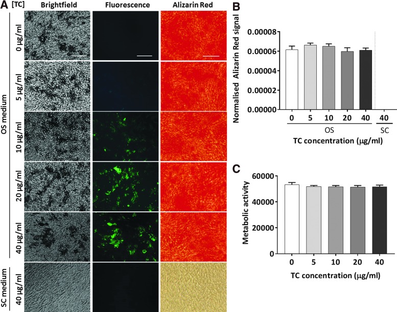
The final stage of in vitro osteogenic differentiation is characterized by the production of mineral deposits containing calcium cations and inorganic phosphates, which populate the extracellular matrix (ECM) surrounding the cell monolayer. Conventional histological techniques for the assessment of mineralization, such as Von Kossa and Alizarin Red S staining, are end point techniques requiring cell fixation. Moreover, in both cases staining quantitation requires dye extraction, which irreversibly alters the ECM conformation and structure, therefore preventing the use of the sample for further analysis. In this study, the use of tetracycline hydrochloride (TC) is proposed for the nondestructive staining, quantitation, and imaging of mineralizing bone-like nodules in live cultures of human bone marrow mesenchymal stem cells cultured under osteogenic conditions. Overnight administration of TC to living cells was shown not to alter the metabolic activity or the progression of cell differentiation. When applied to differentiating cultures, cell exposure to serial doses of TC was found to produce quantifiable fluorescence emission specifically in osteogenic cultures. Incubation with TC enabled fluorescence imaging of mineralized areas in live cultures and the combination with other fluorophores using appropriate filters. These results demonstrate that serial TC administration over the differentiation time course provides a qualitative and quantitative tool for the monitoring and evaluation of the differentiation process in live cells.
体外成骨分化的最后阶段的特征是产生含有钙离子和无机磷酸盐的矿物沉积物,这些沉积物填充细胞单层周围的细胞外基质(ECM)。评估矿化的传统组织学技术,如Von Kossa 和茜素红 S 染色,是需要细胞固定的终点技术。此外,在这两种情况下,染色定量需要染料提取,这会不可逆地改变 ECM 的构象和结构,因此阻止了对样本进行进一步分析。在这项研究中,建议使用盐酸四环素(TC)对活培养的人骨髓间充质干细胞在成骨条件下培养的矿化骨样结节进行非破坏性染色、定量和成像。将 TC overnight 给药于活细胞不会改变细胞代谢活性或细胞分化的进程。当应用于分化培养物时,发现细胞暴露于 TC 的连续剂量会在成骨培养物中产生可定量的荧光发射。TC 的孵育使活培养物中矿化区域的荧光成像成为可能,并且可以与使用适当滤光片的其他荧光团结合使用。这些结果表明,在分化过程中连续 TC 给药提供了一种定性和定量工具,用于监测和评估活细胞中的分化过程。