Program of Stem Cells and Regenerative Medicine, Affiliated Guangzhou Women and Children's Hospital, Zhongshan School of Medicine, Sun Yat-Sen University, Guangzhou, 510080, China.
Center for Stem Cell Biology and Tissue Engineering, Key Laboratory for Stem Cells and Tissue Engineering, Ministry of Education, Sun Yat-Sen University, 74# Zhongshan 2nd Road, Guangzhou, Guangdong, China.
J Hematol Oncol. 2018 Jan 22;11(1):11. doi: 10.1186/s13045-018-0554-z.
Despite the high cure rate of T cell acute lymphoblastic leukemia (T-ALL), drug resistance to chemotherapy remains a significant clinical problem. Bone marrow mesenchymal stem cells (MSCs) protect leukemic cells from chemotherapy, but the underlying mechanisms are poorly understood. In this study, we aimed to uncover the mechanism of MSC-induced chemoresistance in T-ALL cells, thus providing a promising clinical therapy target.
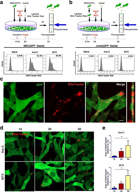
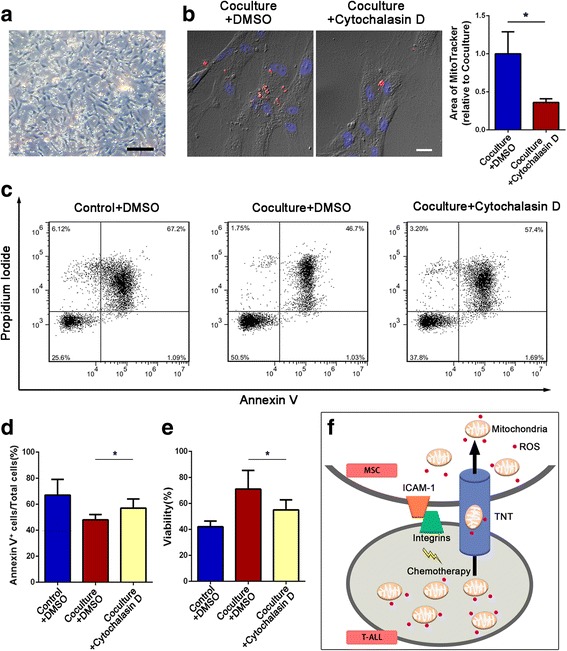
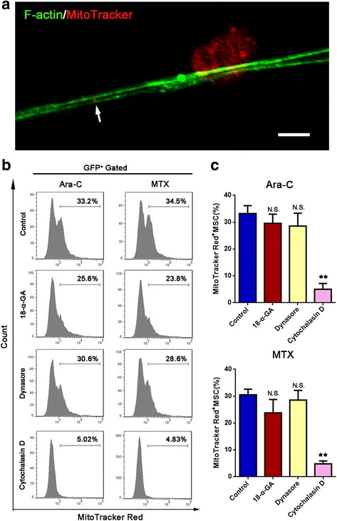
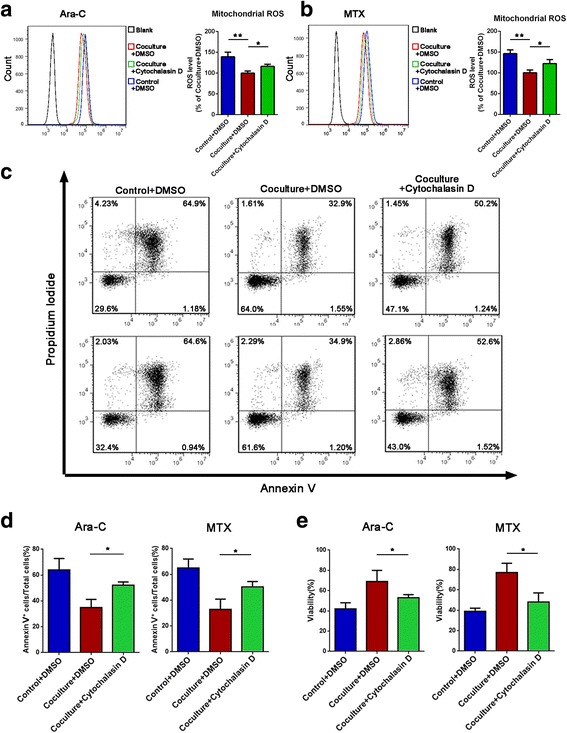
Cell viability was determined using the viability assay kit CCK-8. The mitochondrial ROS levels were detected using the fluorescent probe MitoSOX™ Red, and fluorescence intensity was measured by flow cytometry. In vitro, MSCs and Jurkat cells were cocultured. MSCs were labeled with green fluorescent protein (GFP), and Jurkat cells were labeled with the mitochondria-specific dye MitoTracker Red. Bidirectional mitochondrial transfer was detected by flow cytometry and confocal microscopy. The mechanism of mitochondria transfer was analyzed by inhibitor assays. Transcripts related to Jurkat cell/MSC adhesion in the coculture system were assessed by qRT-PCR. After treatment with a neutralizing antibody against a key adhesion molecule, mitochondria transfer from Jurkat cells to MSCs was again detected by flow cytometry and confocal microscopy. Finally, we verified our findings using human primary T-ALL cells cocultured with MSCs.
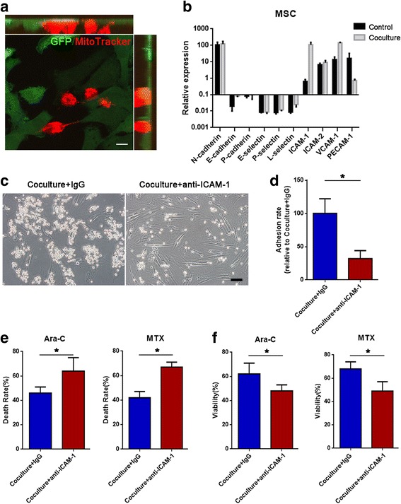
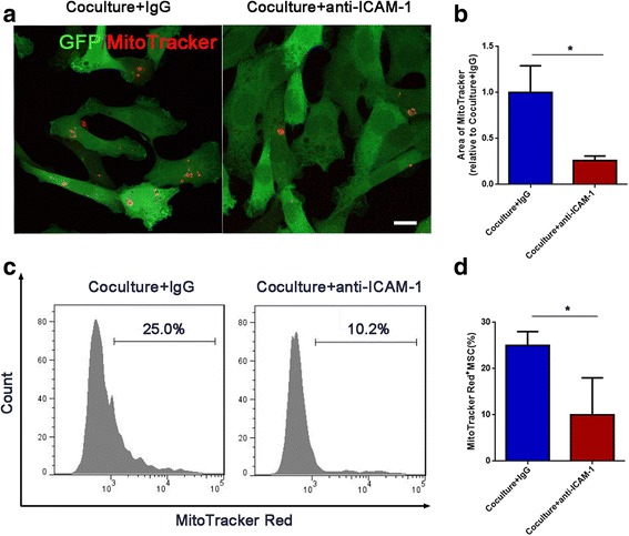
Chemotherapeutic drugs caused intracellular oxidative stress in Jurkat cells. Jurkat cells transfer mitochondria to MSCs but receive few mitochondria from MSCs, resulting in chemoresistance. This process of mitochondria transfer is mediated by tunneling nanotubes, which are protrusions that extend from the cell membrane . Moreover, we found that most Jurkat cells adhered to MSCs in the coculture system, which was mediated by the adhesion molecule ICAM-1. Treatment with a neutralizing antibody against ICAM-1 led to a decreased number of adhering Jurkat cells, decreased mitochondria transfer, and increased chemotherapy-induced cell death.
We show evidence that mitochondria transfer from Jurkat cells to MSCs, which is mediated by cell adhesion, may be a potential therapeutic target for T-ALL treatment.
尽管 T 细胞急性淋巴细胞白血病 (T-ALL) 的治愈率很高,但对化疗的耐药性仍然是一个重大的临床问题。骨髓间充质干细胞 (MSCs) 可保护白血病细胞免受化疗药物的杀伤,但其中的机制尚不清楚。在这项研究中,我们旨在揭示 MSC 诱导 T-ALL 细胞耐药的机制,从而为临床治疗提供有前景的靶点。
使用 CCK-8 细胞活力试剂盒检测细胞活力。使用荧光探针 MitoSOX™ Red 检测线粒体 ROS 水平,并通过流式细胞术测量荧光强度。在体外,将 MSCs 和 Jurkat 细胞共培养。用绿色荧光蛋白 (GFP) 标记 MSCs,用线粒体特异性染料 MitoTracker Red 标记 Jurkat 细胞。通过流式细胞术和共聚焦显微镜检测双向线粒体转移。通过抑制剂实验分析线粒体转移的机制。通过 qRT-PCR 评估共培养体系中与 Jurkat 细胞/MSC 黏附相关的转录本。用中和关键黏附分子的抗体处理后,再次通过流式细胞术和共聚焦显微镜检测 Jurkat 细胞向 MSCs 的线粒体转移。最后,我们使用与 MSCs 共培养的人原发性 T-ALL 细胞验证了我们的发现。
化疗药物在 Jurkat 细胞中引起细胞内氧化应激。Jurkat 细胞将线粒体转移至 MSCs,但从 MSCs 中接收的线粒体较少,从而导致耐药性。这种线粒体转移过程是由隧道纳米管介导的,隧道纳米管是从细胞膜延伸出的突起。此外,我们发现共培养体系中大多数 Jurkat 细胞黏附于 MSCs,这是由黏附分子 ICAM-1 介导的。用中和 ICAM-1 的抗体处理后,黏附的 Jurkat 细胞数量减少,线粒体转移减少,化疗诱导的细胞死亡增加。
我们的研究结果表明,Jurkat 细胞向 MSCs 的线粒体转移可能是 T-ALL 治疗的一个潜在治疗靶点,该过程是由细胞黏附介导的。