Department of Neurosurgery, Suqian First Hospital, Suqian, Jiangsu, China.
Department of Neurosurgery, The Affiliated Hospital of Medical College Qingdao University, Qingdao, Shandong, China.
Cell Death Dis. 2018 Jan 24;9(2):99. doi: 10.1038/s41419-017-0155-8.
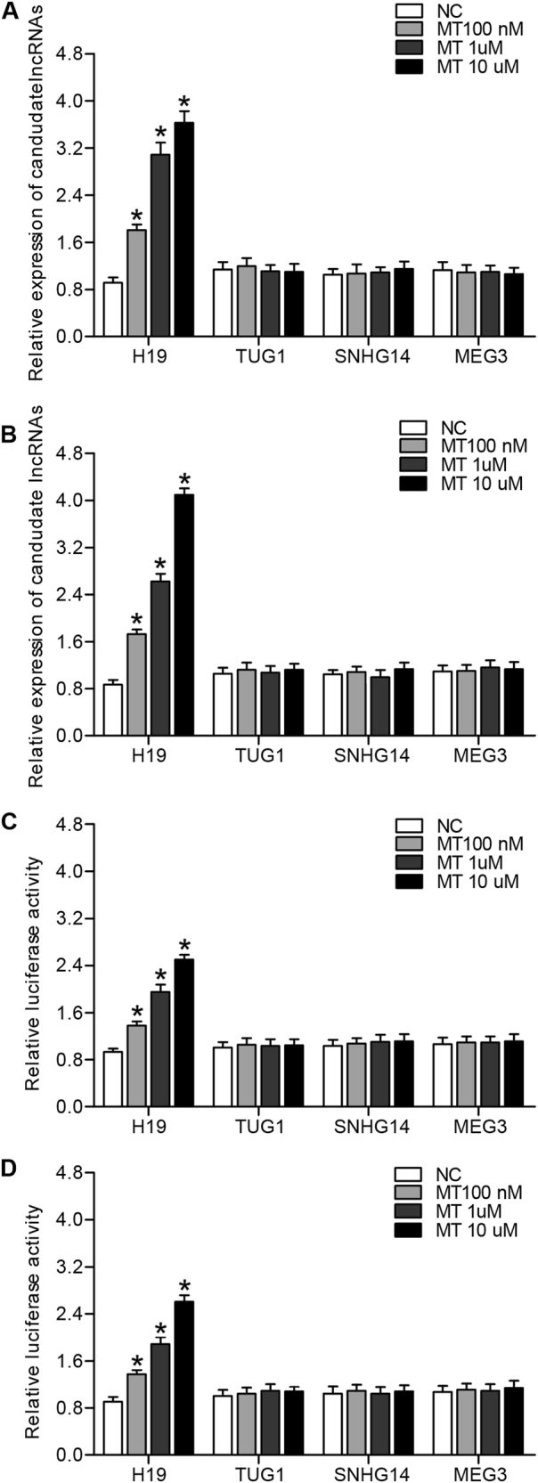
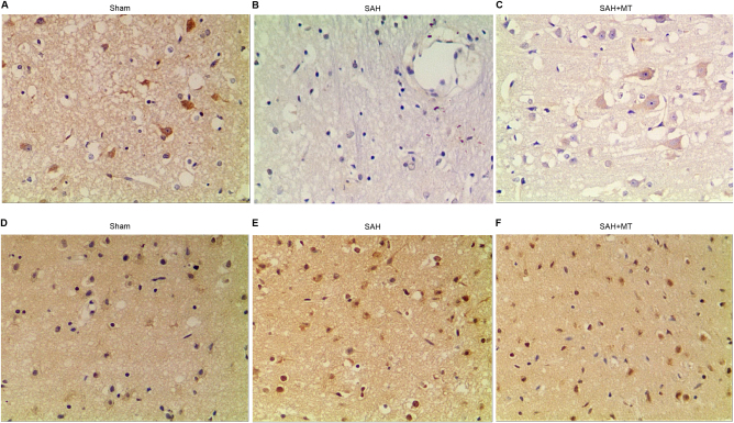
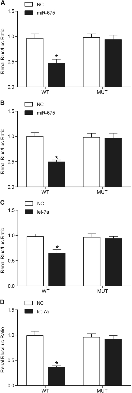
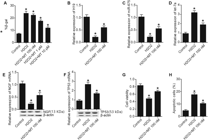
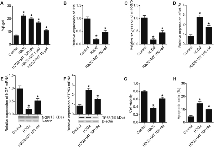
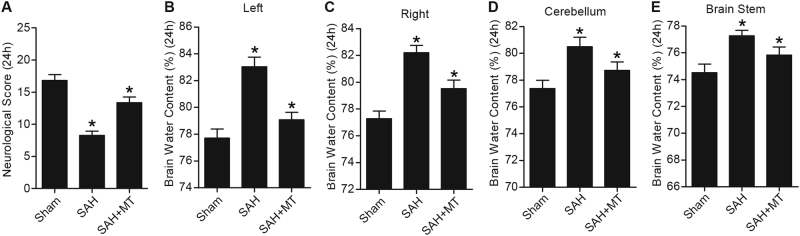
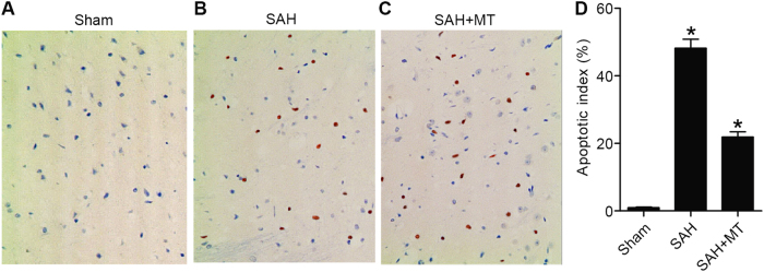
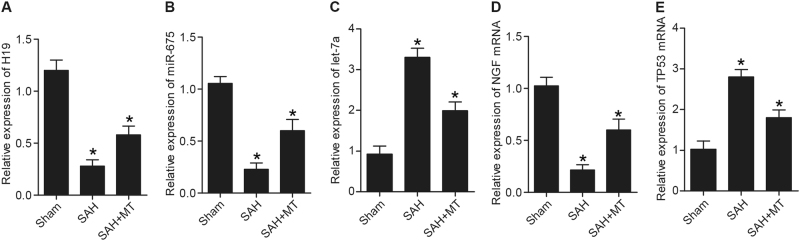
The objective of this study was to identify the protective effect of melatonin (MT) against early brain injury (EBI) following subarachnoid hemorrhage (SAH) and explore the underlying molecular mechanism. Real-time polymerase chain reaction (PCR) and luciferase assay were utilized to detect the effect of MT on H19 expression level, computation analysis and luciferase assay were conducted to the underlying mechanism of let-7a and miR-675. Real-time PCR, western blot analysis, immunohistochemistry, 3-(4,5-dimethylthiazol-2-yl)-2,5-diphenyltetrazolium bromide (MTT) assay, and flow cytometry analysis were performed to detect the effect of MT on H19, miR-675, let-7a, TP53, neural growth factor (NGF) levels, cell viability, and apoptosis status. Melatonin increased H19 expression level by enhancing H19 transcriptional efficiency in a concentration-dependent manner. MiR-675 and let-7a directly targeted P53 and NGF, respectively, and miR-675 reduced luciferase activity of wild-type but not mutant TP53 3'UTR. Meanwhile, let-7a suppressed luciferase activity of wild-type but not mutant NGF 3'UTR. HO increased number of SA-b-gal, and while MT administration repressed the premature senescence. HO obviously upregulated expressions of H19, miR-675, and NGF, and downregulated let-7a and TP53 levels; however, MT treatment reduced expressions of H19, miR-675, and NGF, and improved let-7a and TP53 levels. Treating with MT attenuated the neurological deficits and reduced the brain swelling. MT treatment repressed apoptosis of neurons caused by SAH. Levels of H19, miR-675, and NGF were much higher in the SAH + MT group, while there were even higher levels of H19, miR-675, and NGF in the SAH group than in the sham group; levels of let-7a and TP53 were much lower in the SAH + MT group, while they were even lower in the SAH group than in the sham group. Our study revealed that treatment with MT protected against EBI after SAH by modulating the signaling pathways of H19-miR-675-P53-apoptosis and H19-let-7a-NGF-apoptosis.
本研究旨在探讨褪黑素(MT)对蛛网膜下腔出血(SAH)后早期脑损伤(EBI)的保护作用,并探讨其潜在的分子机制。采用实时聚合酶链反应(PCR)和荧光素酶检测 MT 对 H19 表达水平的影响,通过计算分析和荧光素酶检测潜在的机制 let-7a 和 miR-675。采用实时 PCR、western blot 分析、免疫组织化学、3-(4,5-二甲基噻唑-2-基)-2,5-二苯基四氮唑溴盐(MTT)检测和流式细胞术分析检测 MT 对 H19、miR-675、let-7a、TP53、神经生长因子(NGF)水平、细胞活力和凋亡状态的影响。褪黑素通过增强 H19 转录效率,以浓度依赖的方式增加 H19 的表达水平。miR-675 和 let-7a 分别直接靶向 P53 和 NGF,而 miR-675 降低野生型但不降低突变型 TP53 3'UTR 的荧光素酶活性。同时,let-7a 抑制野生型但不抑制突变型 NGF 3'UTR 的荧光素酶活性。HO 增加了 SA-b-gal 的数量,而 MT 给药抑制了过早衰老。HO 明显上调 H19、miR-675 和 NGF 的表达,下调 let-7a 和 TP53 水平;然而,MT 处理降低了 H19、miR-675 和 NGF 的表达,并改善了 let-7a 和 TP53 的水平。MT 处理减轻了 SAH 引起的神经功能缺损和脑肿胀。MT 处理抑制了 SAH 引起的神经元凋亡。SAH+MT 组 H19、miR-675 和 NGF 水平明显升高,而 SAH 组甚至高于假手术组;SAH+MT 组 let-7a 和 TP53 水平明显降低,而 SAH 组甚至低于假手术组。我们的研究表明,MT 通过调节 H19-miR-675-P53-凋亡和 H19-let-7a-NGF-凋亡信号通路,对 SAH 后 EBI 起到保护作用。