Huang Jun, Yan Zi-Ning, Rui Yi-Fei, Fan Li, Liu Chang, Li Jie
Department of Echocardiography, the Affiliated Changzhou No.2 People's Hospital of Nanjing Medical University, Changzhou, China.
BMC Cardiovasc Disord. 2018 Jan 30;18(1):13. doi: 10.1186/s12872-018-0753-0.
Hypertrophic cardiomyopathy (HCM) is a genetic disease was characterised by left ventricular hypertrophy (LVH), myocardial fibrosis, fiber disarray. The short-axis systolic function is important in left ventricle function.
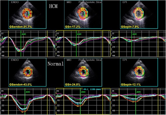
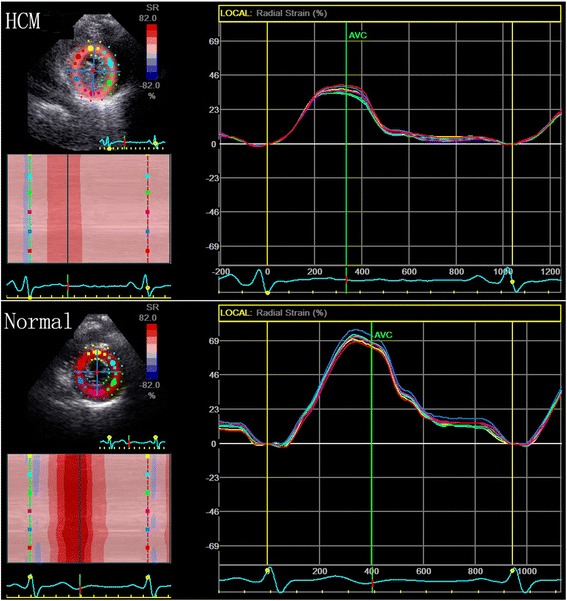
Forty one healthy subjects and 37 HCM patients were enrolled for this research. Parasternal short-axis at the basal, middle, and apical levels were acquired by Echocardiography. The peak systolic circumferential strain of the endocardial, the middle and the epicardial layers, the peak systolic radial strain, and the peak systolic rotational degrees at different short-axis levels were measured by 2-dimensional speckle tracking imaging (2D-STI).
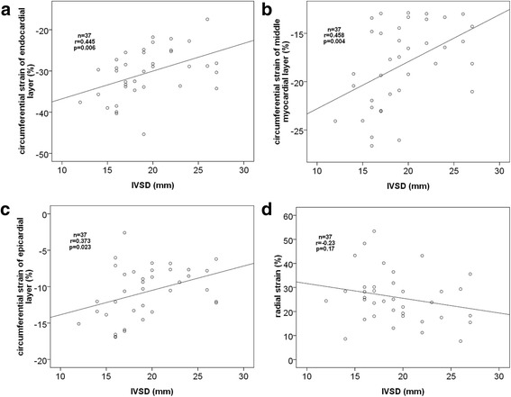
The peak systolic circumferential strain of the septum and anterior walls in HCM patients was significantly lower than normal subjects. All of the peak systolic radial strain in HCM patients was significantly lower than normal subjects. The rotational degrees at the base and middle short-axis levels in HCM patients were larger than normal subjects. The interventricular septal thickness in end-diastolic period correlated to the peak systolic circumferential strain of the septum wall.
The short-axis systolic function was impaired in HCM patients. The peak circumferential systolic strain of the different layers, peak systolic radial strain and rotation degrees of the different short-axis levels detected by 2D-STI are very feasible for assessing the short-axis function in HCM patients.
肥厚型心肌病(HCM)是一种遗传性疾病,其特征为左心室肥厚(LVH)、心肌纤维化、纤维排列紊乱。短轴收缩功能在左心室功能中很重要。
本研究纳入了41名健康受试者和37名HCM患者。通过超声心动图获取心底、心中和心尖水平的胸骨旁短轴图像。采用二维斑点追踪成像(2D-STI)测量不同短轴水平的心内膜、中层和心外膜层的收缩期圆周应变峰值、收缩期径向应变峰值以及收缩期旋转角度峰值。
HCM患者室间隔和前壁的收缩期圆周应变峰值显著低于正常受试者。HCM患者的所有收缩期径向应变峰值均显著低于正常受试者。HCM患者心底和心中短轴水平的旋转角度大于正常受试者。舒张末期室间隔厚度与室间隔壁的收缩期圆周应变峰值相关。
HCM患者的短轴收缩功能受损。二维斑点追踪成像检测到的不同层面的收缩期圆周应变峰值、不同短轴水平的收缩期径向应变峰值和旋转角度对于评估HCM患者的短轴功能非常可行。