Department of Endocrinology, Huai'an Hospital Affiliated to Xuzhou Medical University, and Huai'an Second People's Hospital, Huai'an 223002, Jiangsu, China; Department of Biochemistry and Molecular Biology, The College of Basic Medical Sciences, The Second Military Medical University, Shanghai 200433, China.
Department of Gastrointestinal Surgery/Clinical Nutrition, Beijing Shijitan Hospital, Capital Medical University, Beijing 100038, China.
J Lipid Res. 2018 Apr;59(4):625-634. doi: 10.1194/jlr.M082040. Epub 2018 Feb 5.
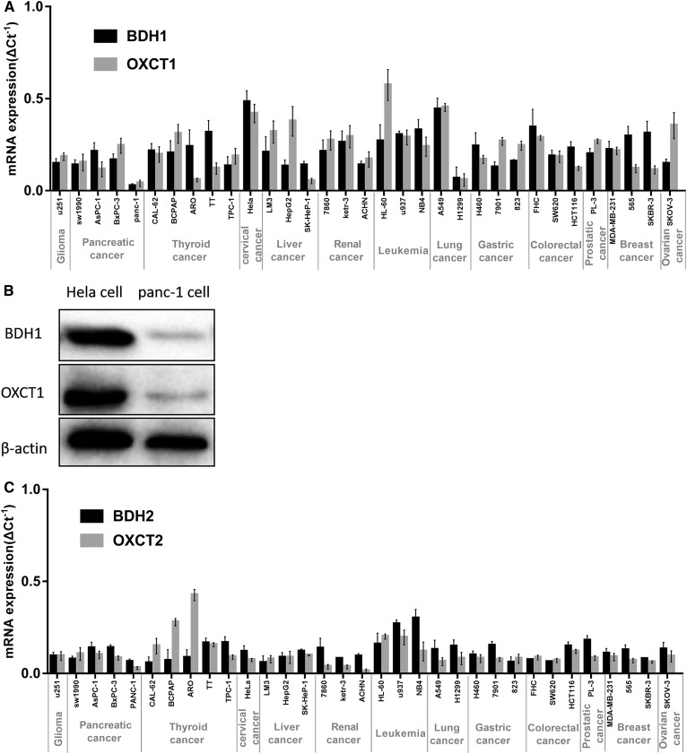
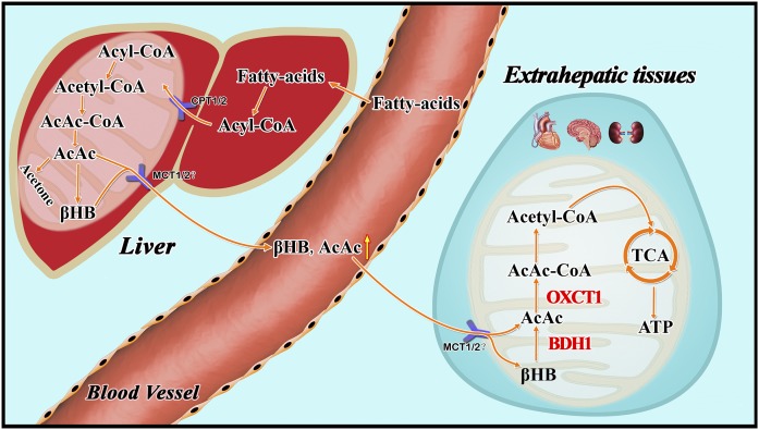
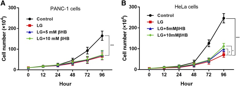
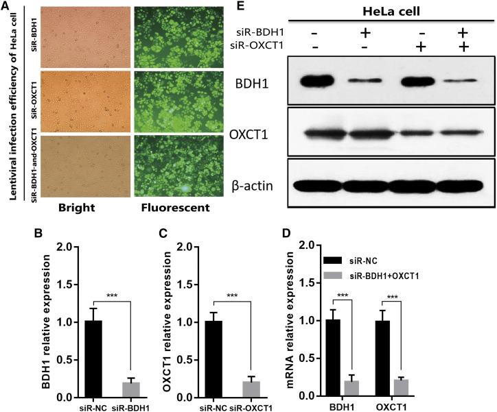
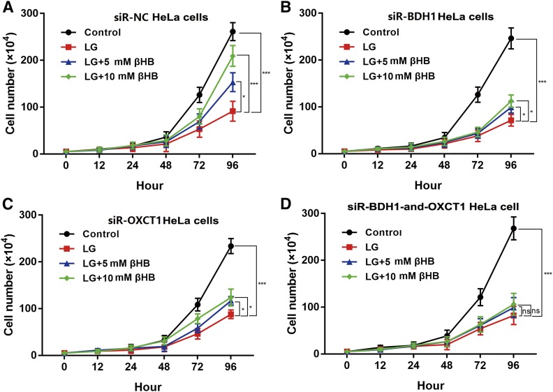
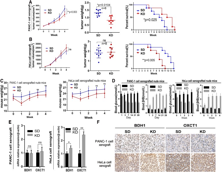
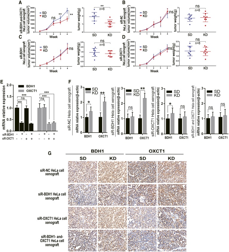
The ketogenic diet (KD) is a high-fat, very-low-carbohydrate diet that triggers a fasting state by decreasing glucose and increasing ketone bodies, such as β-hydroxybutyrate (βHB). In experimental models and clinical trials, the KD has shown anti-tumor effects, possibly by reducing energy supplies to cells, which damage the tumor microenvironment and inhibit tumor growth. Here, we determined expression levels of genes encoding the ketolytic enzymes 3-hydroxybutyrate dehydrogenase 1 (BDH1) and succinyl-CoA: 3-oxoacid CoA transferase 1 (OXCT1) in 33 human cancer cell lines. We then selected two representative lines, HeLa and PANC-1, for in vivo examination of KD sensitivity in tumors with high or low expression, respectively, of these two enzymes. In mice with HeLa xenografts, the KD increased tumor growth and mouse survival decreased, possibly because these tumors actively consumed ketone bodies as an energy source. Conversely, the KD significantly inhibited growth of PANC-1 xenograft tumors. βHB added to each cell culture significantly increased proliferation of HeLa cells, but not PANCI-1 cells. Downregulation of both BDH1 and OXCT1 rendered HeLa cells sensitive to the KD in vitro and in vivo. Tumors with low ketolytic enzyme expression may be unable to metabolize ketone bodies, thus predicting a better response to KD therapy.
生酮饮食(KD)是一种高脂肪、低碳水化合物的饮食,通过降低葡萄糖和增加酮体(如β-羟丁酸(βHB))来模拟禁食状态。在实验模型和临床试验中,KD 显示出抗肿瘤作用,可能是通过减少细胞的能量供应来破坏肿瘤微环境并抑制肿瘤生长。在这里,我们测定了 33 个人类癌细胞系中编码酮解酶 3-羟丁酸脱氢酶 1(BDH1)和琥珀酰辅酶 A:3-氧代酸辅酶 A 转移酶 1(OXCT1)的基因表达水平。然后,我们选择了两个代表性的细胞系,HeLa 和 PANC-1,分别用于研究这两种酶高表达和低表达的肿瘤对 KD 敏感性的体内研究。在 HeLa 异种移植小鼠中,KD 增加了肿瘤生长,降低了小鼠存活率,这可能是因为这些肿瘤积极地将酮体作为能量来源消耗。相反,KD 显著抑制了 PANC-1 异种移植肿瘤的生长。βHB 加入每种细胞培养物中显著增加了 HeLa 细胞的增殖,但不增加 PANC-1 细胞的增殖。BDH1 和 OXCT1 的下调使 HeLa 细胞在体外和体内对 KD 敏感。低酮解酶表达的肿瘤可能无法代谢酮体,因此预测对 KD 治疗有更好的反应。