X-ray Science Division, Argonne National Laboratory, 9700 South Cass Avenue, Lemont, IL, 60439, USA.
Argonne Leadership Computing Facility (ALCF), Argonne National Laboratory, 9700 South Cass Avenue, Lemont, IL, 60439, USA.
Sci Rep. 2018 Feb 7;8(1):2575. doi: 10.1038/s41598-018-19426-7.
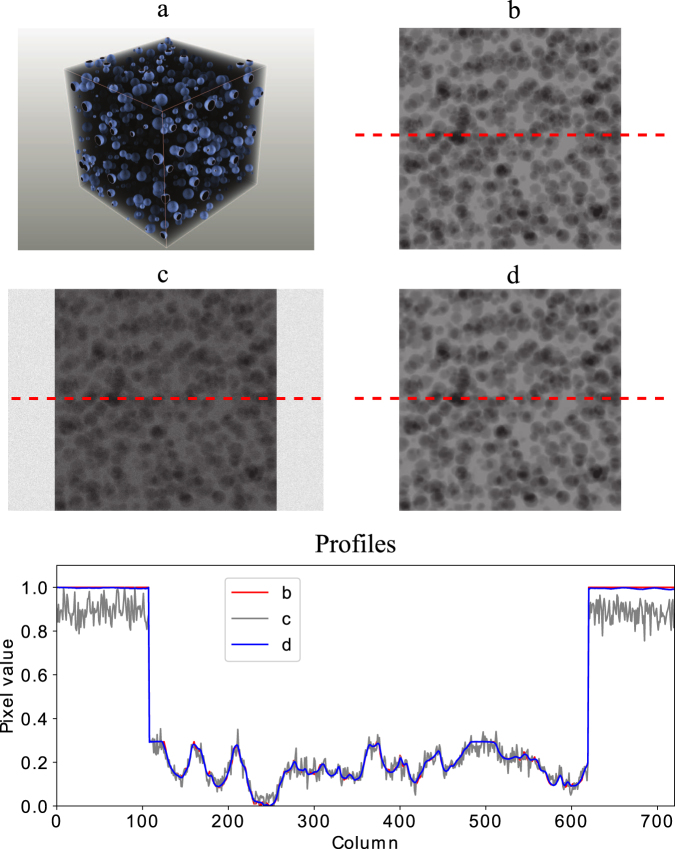
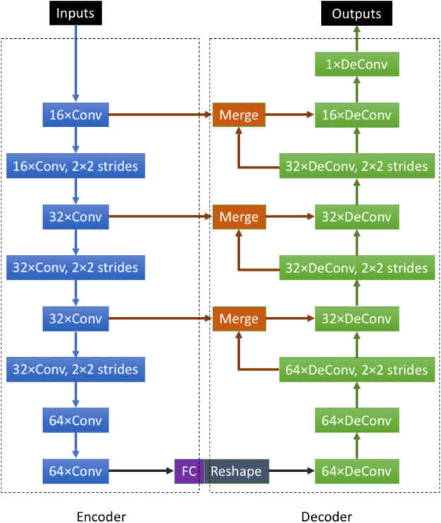
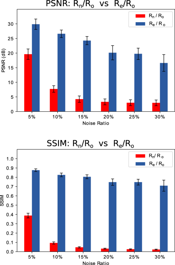
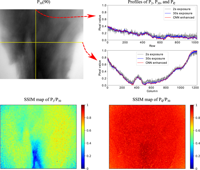
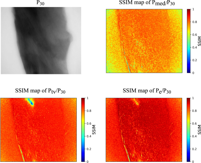
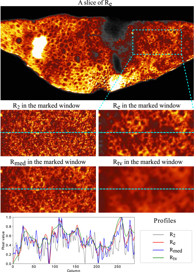
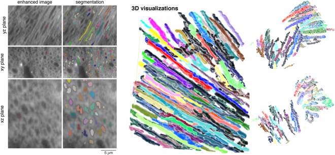
Synchrotron-based X-ray tomography offers the potential for rapid large-scale reconstructions of the interiors of materials and biological tissue at fine resolution. However, for radiation sensitive samples, there remain fundamental trade-offs between damaging samples during longer acquisition times and reducing signals with shorter acquisition times. We present a deep convolutional neural network (CNN) method that increases the acquired X-ray tomographic signal by at least a factor of 10 during low-dose fast acquisition by improving the quality of recorded projections. Short-exposure-time projections enhanced with CNNs show signal-to-noise ratios similar to long-exposure-time projections. They also show lower noise and more structural information than low-dose short-exposure acquisitions post-processed by other techniques. We evaluated this approach using simulated samples and further validated it with experimental data from radiation sensitive mouse brains acquired in a tomographic setting with transmission X-ray microscopy. We demonstrate that automated algorithms can reliably trace brain structures in low-dose datasets enhanced with CNN. This method can be applied to other tomographic or scanning based X-ray imaging techniques and has great potential for studying faster dynamics in specimens.
基于同步加速器的 X 射线断层扫描技术具有在精细分辨率下快速重建材料和生物组织内部的潜力。然而,对于对辐射敏感的样品,在更长的采集时间内损坏样品和在更短的采集时间内减少信号之间仍然存在基本的权衡。我们提出了一种深度卷积神经网络(CNN)方法,该方法通过提高记录投影的质量,在低剂量快速采集期间将采集的 X 射线断层扫描信号提高至少 10 倍。经过 CNN 增强的短曝光时间投影显示出与长曝光时间投影相似的信噪比。与其他技术后处理的低剂量短曝光采集相比,它们还显示出更低的噪声和更多的结构信息。我们使用模拟样本评估了这种方法,并进一步使用在层析 X 射线显微镜的透射设置下从对辐射敏感的老鼠大脑获得的实验数据进行了验证。我们证明了自动化算法可以可靠地追踪经过 CNN 增强的低剂量数据集的大脑结构。这种方法可以应用于其他层析或基于扫描的 X 射线成像技术,并且在研究样本中更快的动态方面具有很大的潜力。