Department of Respiratory Medicine, Academic Medical Centre, Meibergdreef 9, Amsterdam, The Netherlands.
Respir Res. 2011 Jun 23;12(1):85. doi: 10.1186/1465-9921-12-85.
Airway remodelling is a feature of asthma including fragmentation of elastic fibres observed in the superficial elastin network of the airway wall. Fibered confocal fluorescence microscopy (FCFM) is a new and non-invasive imaging technique performed during bronchoscopy that may visualize elastic fibres, as shown by in vitro spectral analysis of elastin powder. We hypothesized that FCFM images capture in vivo elastic fibre patterns within the airway wall and that such patterns correspond with airway histology. We aimed to establish the concordance between the bronchial elastic fibre pattern in histology and FCFM. Second, we examined whether elastic fibre patterns in histology and FCFM were different between asthmatic subjects and healthy controls. Finally, the association between these patterns and lung function parameters was investigated.
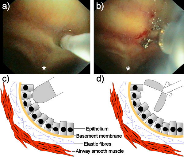
In a cross-sectional study comprising 16 subjects (8 atopic asthmatic patients with controlled disease and 8 healthy controls) spirometry and bronchoscopy were performed, with recording of FCFM images followed by endobronchial biopsy at the airway main carina. Elastic fibre patterns in histological sections and FCFM images were scored semi-quantitatively. Agreement between histology and FCFM was analysed using linearly weighted kappa κw.
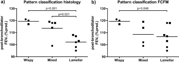
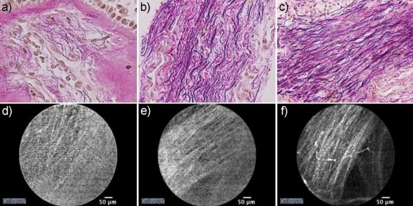
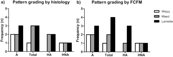
The patterns observed in histological sections and FCFM images could be divided into 3 distinct groups. There was good agreement between elastic fibre patterns in histology and FCFM patterns (κw 0.744). The semi-quantitative pattern scores were not different between asthmatic patients and controls. Notably, there was a significant difference in post-bronchodilator FEV1 %predicted between the different patterns by histology (p = 0.001) and FCFM (p = 0.048), regardless of asthma or atopy.
FCFM captures the elastic fibre pattern within the airway wall in humans in vivo. The association between post-bronchodilator FEV1 %predicted and both histological and FCFM elastic fibre patterns points towards a structure-function relationship between extracellular matrix in the airway wall and lung function.
Netherlands Trial Register NTR1306.
气道重塑是哮喘的一个特征,包括气道壁浅层弹力网络中弹性纤维的断裂。纤维状共聚焦荧光显微镜(FCFM)是一种新的非侵入性成像技术,可在支气管镜检查期间进行,通过对弹力蛋白粉末的体外光谱分析,可能显示弹性纤维。我们假设 FCFM 图像可以捕获气道壁内的弹性纤维的体内模式,并且这些模式与气道组织学相对应。我们旨在确定组织学中支气管弹性纤维模式与 FCFM 之间的一致性。其次,我们检查了组织学和 FCFM 中弹性纤维模式在哮喘患者和健康对照者之间是否不同。最后,研究了这些模式与肺功能参数之间的关系。
在一项包含 16 名受试者(8 名特应性哮喘患者和 8 名健康对照者)的横断面研究中,进行了肺活量测定和支气管镜检查,记录 FCFM 图像,然后在气道主隆突处进行支气管内活检。对组织学切片和 FCFM 图像中的弹性纤维模式进行半定量评分。使用线性加权 kappa κw 分析组织学和 FCFM 之间的一致性。
在组织学切片和 FCFM 图像中观察到的模式可以分为 3 个不同的组。组织学和 FCFM 模式之间的一致性很好(κw 0.744)。哮喘患者和对照组之间的半定量模式评分没有差异。值得注意的是,组织学和 FCFM 不同模式之间的支气管扩张剂后 FEV1 %预计值存在显著差异(p = 0.001 和 p = 0.048),无论是否患有哮喘或特应性。
FCFM 在体内捕获了人类气道壁内的弹性纤维模式。支气管扩张剂后 FEV1 %预计值与组织学和 FCFM 弹性纤维模式之间的关联表明,气道壁细胞外基质与肺功能之间存在结构-功能关系。
荷兰试验注册 NTR1306。