Donnelly Brooke, Touyz Stephen, Hay Phillipa, Burton Amy, Russell Janice, Caterson Ian
1School of Psychology, Clinical Psychology Unit, University of Sydney, Sydney,, New South Wales Australia.
2Translational Health Research Institute (THRI), School of Medicine, Western Sydney University, Sydney, New South Wales Australia.
J Eat Disord. 2018 Feb 20;6:3. doi: 10.1186/s40337-018-0187-1. eCollection 2018.
In recent decades there has been growing interest in the use of neuroimaging techniques to explore the structural and functional brain changes that take place in those with eating disorders. However, to date, the majority of research has focused on patients with anorexia nervosa. This systematic review addresses a gap in the literature by providing an examination of the published literature on the neurobiology of individuals who binge eat; specifically, individuals with bulimia nervosa (BN) and binge eating disorder (BED).
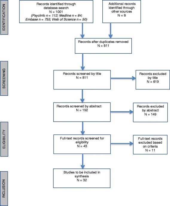
A systematic review was conducted in accordance with PRISMA guidelines using PubMed, PsycInfo, Medline and Web of Science, and additional hand searches through reference lists. 1,003 papers were identified in the database search. Published studies were included if they were an original research paper written in English; studied humans only; used samples of participants with a diagnosed eating disorder characterised by recurrent binge eating; included a healthy control sample; and reported group comparisons between clinical groups and healthy control groups.
Thirty-two papers were included in the systematic review. Significant heterogeneity in the methods used in the included papers coupled with small sample sizes impeded the interpretation of results. Twenty-one papers utilised functional Magnetic Resonance Imaging (fMRI); seven papers utilized Magnetic Resonance Imaging (MRI) with one of these using both MRI and Positron Emission Technology (PET); three studies used Single-Photon Emission Computed Tomography (SPECT) and one study used PET only. A small number of consistent findings emerged in individuals in the acute phase of illness with BN or BED including: volume reduction and increases across a range of areas; hypoactivity in the frontostriatal circuits; and aberrant responses in the insula, amygdala, middle frontal gyrus and occipital cortex to a range of different stimuli or tasks; a link between illness severity in BN and neural changes; diminished attentional capacity and early learning; and in SPECT studies, increased rCBF in relation to disorder-related stimuli.
Studies included in this review are heterogenous, preventing many robust conclusions from being drawn. The precise neurobiology of BN and BED remains unclear and ongoing, large-scale investigations are required. One clear finding is that illness severity, exclusively defined as the frequency of binge eating or bulimic episodes, is related to greater neural changes. The results of this review indicate additional research is required, particularly extending findings of reduced cortical volumes and diminished activity in regions associated with self-regulation (frontostriatal circuits) and further exploring responses to disorder-related stimuli in people with BN and BED.
近几十年来,人们越来越有兴趣使用神经成像技术来探索饮食失调者大脑的结构和功能变化。然而,迄今为止,大多数研究都集中在神经性厌食症患者身上。本系统评价通过对已发表的关于暴饮暴食个体神经生物学的文献进行考察,填补了这一文献空白;具体而言,是针对神经性贪食症(BN)和暴饮暴食症(BED)患者。
按照PRISMA指南,通过PubMed、PsycInfo、Medline和Web of Science进行系统评价,并通过参考文献列表进行额外的手工检索。在数据库搜索中识别出1003篇论文。纳入的已发表研究需满足以下条件:为用英文撰写的原创研究论文;仅研究人类;使用被诊断为以反复暴饮暴食为特征的饮食失调参与者样本;包括健康对照样本;并报告临床组与健康对照组之间的组间比较。
32篇论文被纳入系统评价。纳入论文中使用的方法存在显著异质性,且样本量较小,妨碍了结果的解释。21篇论文使用功能磁共振成像(fMRI);7篇论文使用磁共振成像(MRI),其中1篇同时使用了MRI和正电子发射断层扫描技术(PET);3项研究使用单光子发射计算机断层扫描(SPECT),1项研究仅使用PET。在BN或BED急性期个体中出现了一些一致的发现,包括:多个区域的体积减少和增加;额纹状体回路活动减退;岛叶、杏仁核、额中回和枕叶皮质对一系列不同刺激或任务的异常反应;BN疾病严重程度与神经变化之间的联系;注意力容量和早期学习能力下降;以及在SPECT研究中,与疾病相关刺激有关的局部脑血流增加。
本综述纳入的研究具有异质性,无法得出许多确凿的结论。BN和BED的确切神经生物学机制仍不清楚,需要进行持续的大规模研究。一个明确的发现是,仅定义为暴饮暴食或贪食发作频率的疾病严重程度与更大的神经变化有关。本综述结果表明需要进行更多研究,特别是扩展关于皮质体积减小以及与自我调节相关区域(额纹状体回路)活动减退的发现,并进一步探索BN和BED患者对疾病相关刺激的反应。