Institute of Human Genetics, Polish Academy of Sciences, Poznan, Poland.
Faculty of Chemistry, Adam Mickiewicz University, Poznan, Poland.
Sci Rep. 2018 Feb 27;8(1):3682. doi: 10.1038/s41598-018-22018-0.
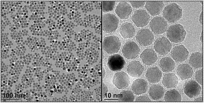
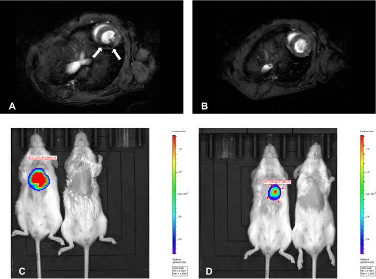
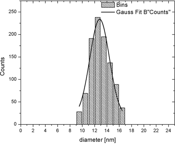
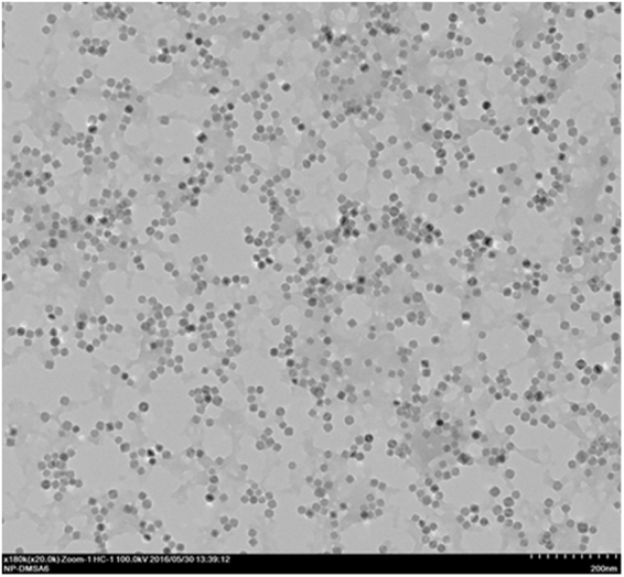
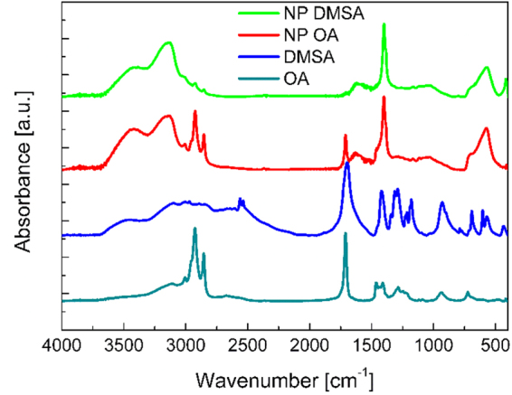
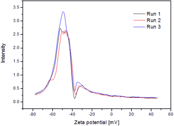
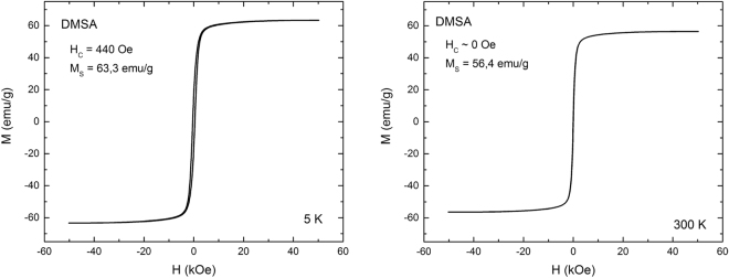
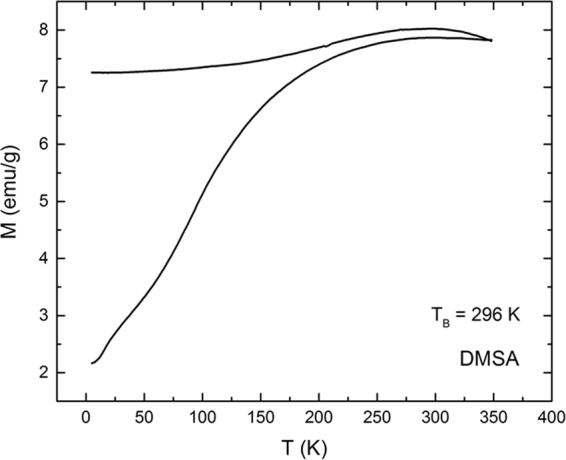
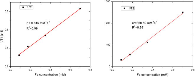
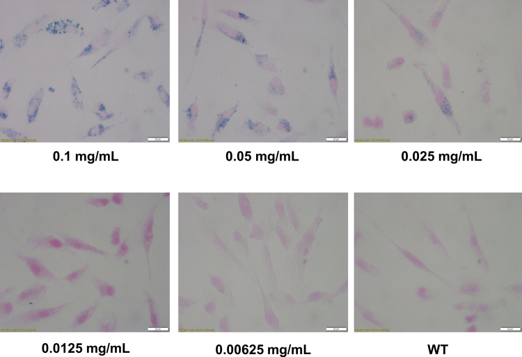
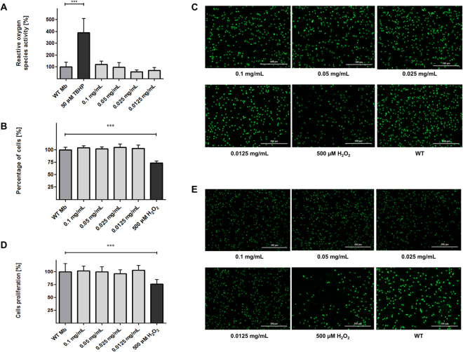
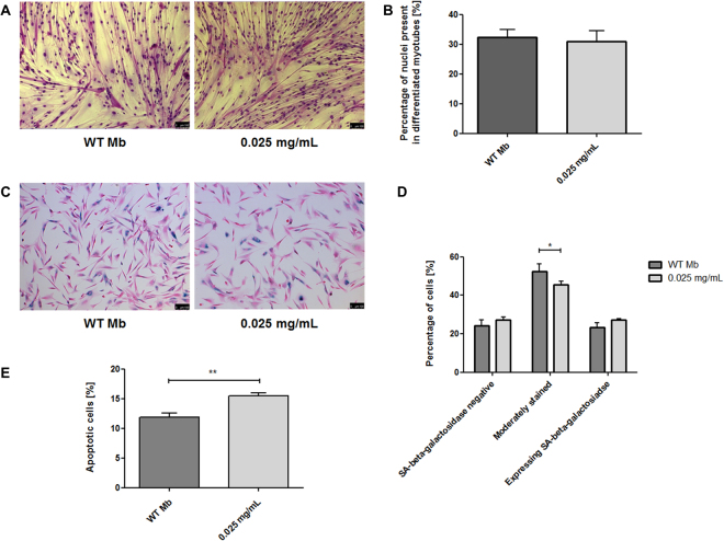
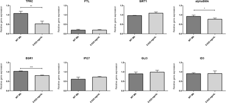
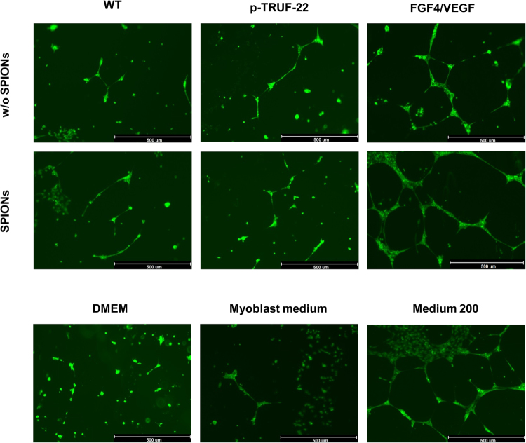
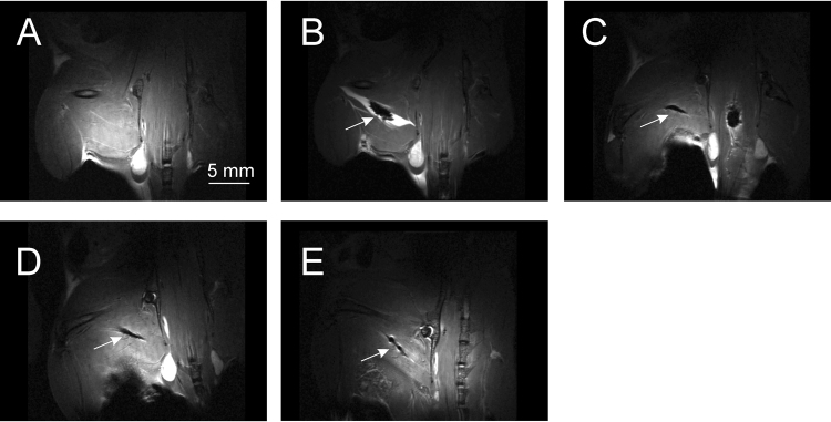
Myocardial infarction (MI) is one of the most frequent causes of death in industrialized countries. Stem cells therapy seems to be very promising for regenerative medicine. Skeletal myoblasts transplantation into postinfarction scar has been shown to be effective in the failing heart but shows limitations such, e.g. cell retention and survival. We synthesized and investigated superparamagnetic iron oxide nanoparticles (SPIONs) as an agent for direct cell labeling, which can be used for stem cells imaging. High quality, monodisperse and biocompatible DMSA-coated SPIONs were obtained with thermal decomposition and subsequent ligand exchange reaction. SPIONs' presence within myoblasts was confirmed by Prussian Blue staining and inductively coupled plasma mass spectrometry (ICP-MS). SPIONs' influence on tested cells was studied by their proliferation, ageing, differentiation potential and ROS production. Cytotoxicity of obtained nanoparticles and myoblast associated apoptosis were also tested, as well as iron-related and coating-related genes expression. We examined SPIONs' impact on overexpression of two pro-angiogenic factors introduced via myoblast electroporation method. Proposed SPION-labeling was sufficient to visualize firefly luciferase-modified and SPION-labeled cells with magnetic resonance imaging (MRI) combined with bioluminescence imaging (BLI) in vivo. The obtained results demonstrated a limited SPIONs' influence on treated skeletal myoblasts, not interfering with basic cell functions.
心肌梗死(MI)是工业化国家最常见的死亡原因之一。干细胞疗法似乎非常有前途的再生医学。成肌细胞移植到梗死后的瘢痕组织已被证明对衰竭的心脏有效,但显示出一些局限性,例如细胞保留和存活。我们合成并研究了超顺磁氧化铁纳米粒子(SPIONs)作为直接细胞标记物,可用于干细胞成像。通过热分解和随后的配体交换反应,获得了高质量、单分散和生物相容的 DMSA 包覆的 SPIONs。普鲁士蓝染色和电感耦合等离子体质谱(ICP-MS)证实了 SPIONs 存在于成肌细胞内。通过细胞增殖、衰老、分化潜力和 ROS 产生研究了 SPIONs 对测试细胞的影响。还测试了获得的纳米粒子的细胞毒性和成肌细胞相关的细胞凋亡,以及铁相关和涂层相关基因的表达。我们研究了 SPIONs 对通过成肌细胞电穿孔方法引入的两种促血管生成因子过表达的影响。提出的 SPION 标记足以通过磁共振成像(MRI)结合体内生物发光成像(BLI)可视化萤火虫荧光素酶修饰和 SPION 标记的细胞。所得结果表明 SPIONs 对处理后的成肌细胞的影响有限,不会干扰基本细胞功能。