文献检索文档翻译深度研究
Suppr Zotero 插件Zotero 插件
邀请有礼套餐&价格历史记录

新学期,新优惠

限时优惠:9月1日-9月22日

30天高级会员仅需29元

1天体验卡首发特惠仅需5.99元

了解详情
不再提醒
插件&应用
Suppr Zotero 插件Zotero 插件浏览器插件Mac 客户端Windows 客户端微信小程序
高级版
套餐订阅购买积分包
AI 工具
文献检索文档翻译深度研究
关于我们
关于 Suppr公司介绍联系我们用户协议隐私条款
关注我们

Suppr 超能文献

核心技术专利:CN118964589B侵权必究
粤ICP备2023148730 号-1Suppr @ 2025

超顺磁性氧化铁纳米颗粒对头颈部癌细胞进行磁粒子成像的生物学影响。

Biological impact of superparamagnetic iron oxide nanoparticles for magnetic particle imaging of head and neck cancer cells.

作者信息

Lindemann Antje, Lüdtke-Buzug Kerstin, Fräderich Bianca M, Gräfe Ksenija, Pries Ralph, Wollenberg Barbara

机构信息

Department of Otorhinolaryngology, University Hospital of Schleswig-Holstein, Luebeck, Germany.

Institute of Medical Engineering, University of Luebeck, Luebeck, Germany.

出版信息

Int J Nanomedicine. 2014 Oct 29;9:5025-40. doi: 10.2147/IJN.S63873. eCollection 2014.


DOI:10.2147/IJN.S63873
PMID:25378928
原文链接:https://pmc.ncbi.nlm.nih.gov/articles/PMC4218924/
Abstract

BACKGROUND: 
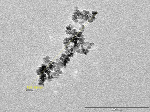
As a tomographic imaging technology, magnetic particle imaging (MPI) allows high spatial resolution and sensitivity, and the possibility to create real-time images by determining the spatial distribution of magnetic particles. To ensure a prospective biosafe application of UL-D (University of Luebeck-Dextran coated superparamagnetic nanoparticles), we evaluated the biocompatibility of superparamagnetic iron oxide nanoparticles (SPIONs), their impact on biological properties, and their cellular uptake using head and neck squamous cancer cells (HNSCCs). METHODS: 
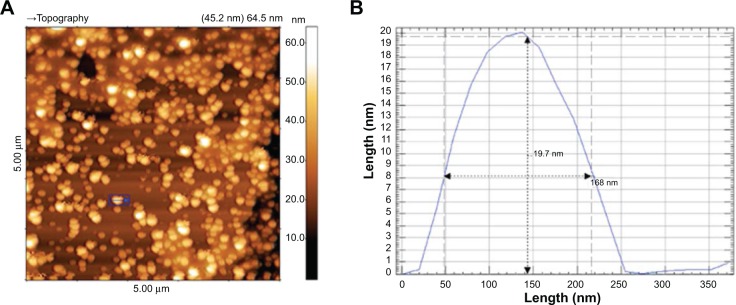
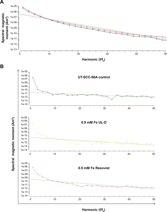
SPIONs that met specific MPI requirements were synthesized as tracers. Labeling and uptake efficiency were analyzed by hematoxylin and eosin staining and magnetic particle spectrometry. Flow cytometry, 3-(4,5-Dimethylthiazol-2-yl)-2,5-diphenyl tetrazolium bromide (MTT) assays, and real-time cell analyzer assays were used to investigate apoptosis, proliferation, and the cytokine response of SPION-labeled cells. The production of reactive oxygen species (ROS) was determined using a fluorescent dye. Experimental results were compared to the contrast agent Resovist(®), a standard agent used in MPI. RESULTS: 
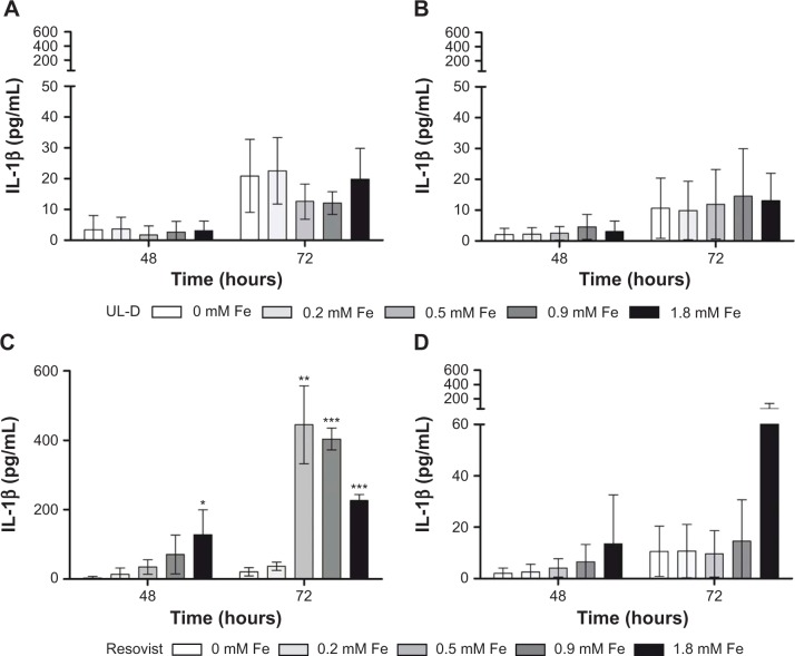
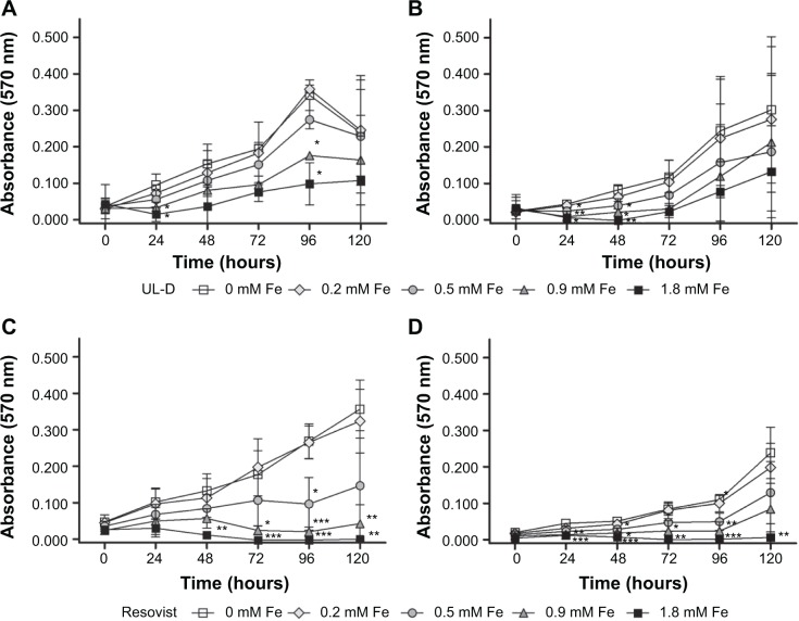
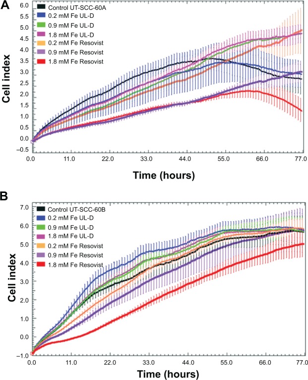
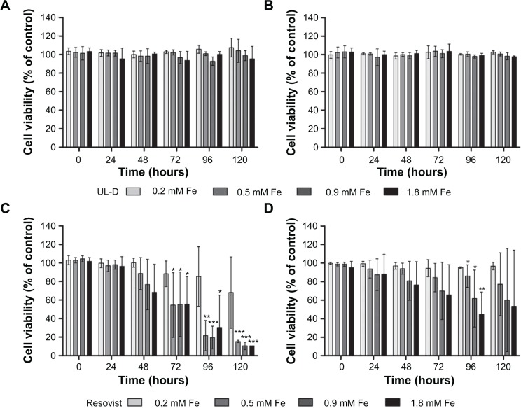
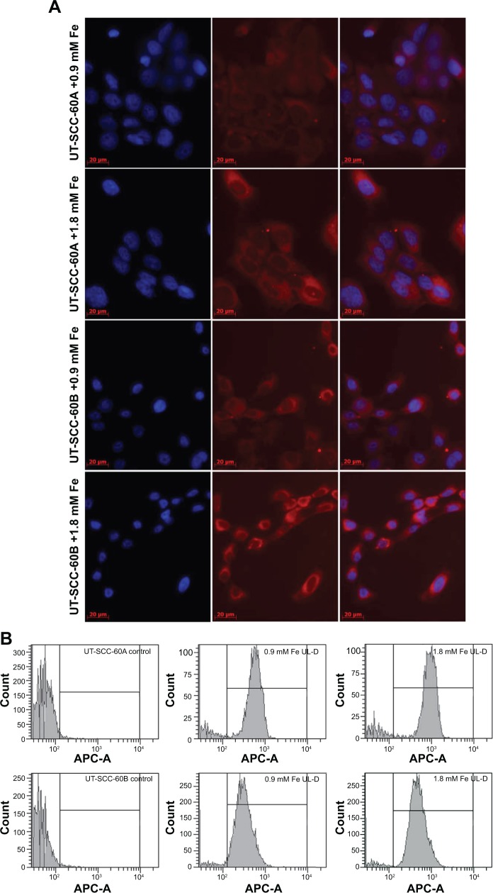
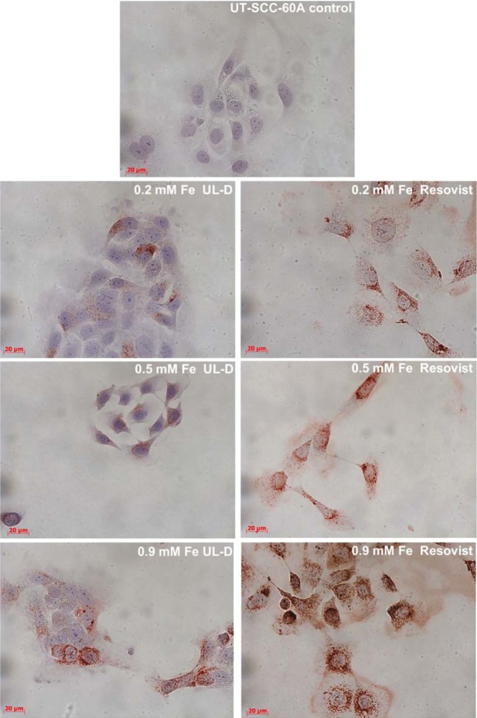
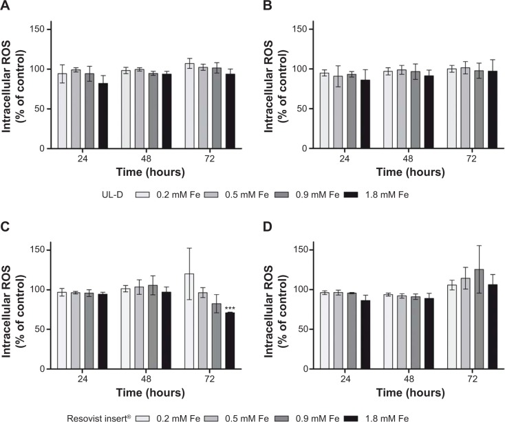
UL-D nanoparticles and Resovist particles were taken up in vitro by HNSCCs via unspecific phagocytosis followed by cytosolic accumulation. To evaluate toxicity, flow cytometry analysis was performed; results showed that dose- and time-dependent administration of Resovist induced apoptosis whereas cell viability of UL-D-labeled cells was not altered. We observed decreased cell proliferation in response to increased SPION concentrations. An intracellular production of ROS could not be detected, suggesting that the particles did not cause oxidative stress. Tumor necrosis factor alpha (TNF-α) and interleukins IL-6, IL-8, and IL-1β were measured to distinguish inflammatory responses. Only the primary tumor cell line labeled with >0.5 mM Resovist showed a significant increase in IL-1β secretion. CONCLUSION: Our data suggest that UL-D SPIONs are a promising tracer material for use in innovative tumor cell analysis in MPI.

摘要

背景:作为一种断层成像技术,磁粒子成像(MPI)具有高空间分辨率和灵敏度,并且能够通过确定磁粒子的空间分布来创建实时图像。为确保超顺磁性氧化铁纳米颗粒(SPIONs)标记的超低剂量右旋糖酐(UL-D,吕贝克大学-右旋糖酐包被的超顺磁性纳米颗粒)在生物安全方面的前瞻性应用,我们使用头颈部鳞状细胞癌(HNSCCs)评估了超顺磁性氧化铁纳米颗粒的生物相容性、其对生物学特性的影响以及细胞摄取情况。 方法:合成符合特定MPI要求的SPIONs作为示踪剂。通过苏木精-伊红染色和磁粒子光谱法分析标记和摄取效率。使用流式细胞术、3-(4,5-二甲基噻唑-2-基)-2,5-二苯基四氮唑溴盐(MTT)测定法和实时细胞分析仪测定法研究SPION标记细胞的凋亡、增殖和细胞因子反应。使用荧光染料测定活性氧(ROS)的产生。将实验结果与MPI中使用的标准造影剂Resovist(®)进行比较。 结果:UL-D纳米颗粒和Resovist颗粒在体外通过非特异性吞噬作用被HNSCCs摄取,随后在细胞质中积累。为评估毒性,进行了流式细胞术分析;结果表明,Resovist的剂量和时间依赖性给药诱导了细胞凋亡,而UL-D标记细胞的活力未改变。我们观察到随着SPION浓度增加,细胞增殖减少。未检测到细胞内ROS的产生,表明这些颗粒未引起氧化应激。测量肿瘤坏死因子α(TNF-α)和白细胞介素IL-6、IL-8和IL-1β以区分炎症反应。只有用>0.5 mM Resovist标记的原发性肿瘤细胞系显示IL-1β分泌显著增加。 结论:我们的数据表明,UL-D SPIONs是一种有前景的示踪剂材料,可用于MPI中创新的肿瘤细胞分析。

https://cdn.ncbi.nlm.nih.gov/pmc/blobs/7ba3/4218924/6c6124c05029/ijn-9-5025Fig11.jpg
https://cdn.ncbi.nlm.nih.gov/pmc/blobs/7ba3/4218924/fef9559d24c7/ijn-9-5025Fig1.jpg
https://cdn.ncbi.nlm.nih.gov/pmc/blobs/7ba3/4218924/ef788f539332/ijn-9-5025Fig2.jpg
https://cdn.ncbi.nlm.nih.gov/pmc/blobs/7ba3/4218924/46e743e6a798/ijn-9-5025Fig3.jpg
https://cdn.ncbi.nlm.nih.gov/pmc/blobs/7ba3/4218924/d3b9b06cdf2e/ijn-9-5025Fig4.jpg
https://cdn.ncbi.nlm.nih.gov/pmc/blobs/7ba3/4218924/2d35c92f75a1/ijn-9-5025Fig5.jpg
https://cdn.ncbi.nlm.nih.gov/pmc/blobs/7ba3/4218924/6cfc6ab02359/ijn-9-5025Fig6.jpg
https://cdn.ncbi.nlm.nih.gov/pmc/blobs/7ba3/4218924/5adfa1fcf721/ijn-9-5025Fig7.jpg
https://cdn.ncbi.nlm.nih.gov/pmc/blobs/7ba3/4218924/5c11f9efb2de/ijn-9-5025Fig8.jpg
https://cdn.ncbi.nlm.nih.gov/pmc/blobs/7ba3/4218924/c26a9f4f7bc3/ijn-9-5025Fig9.jpg
https://cdn.ncbi.nlm.nih.gov/pmc/blobs/7ba3/4218924/be6fe86ffa8f/ijn-9-5025Fig10.jpg
https://cdn.ncbi.nlm.nih.gov/pmc/blobs/7ba3/4218924/6c6124c05029/ijn-9-5025Fig11.jpg

相似文献

[1]
Biological impact of superparamagnetic iron oxide nanoparticles for magnetic particle imaging of head and neck cancer cells.

Int J Nanomedicine. 2014-10-29

[2]
Selection of potential iron oxide nanoparticles for breast cancer treatment based on in vitro cytotoxicity and cellular uptake.

Int J Nanomedicine. 2017-4-19

[3]
Size-dependent superparamagnetic iron oxide nanoparticles dictate interleukin-1β release from mouse bone marrow-derived macrophages.

J Appl Toxicol. 2018-2-28

[4]
Potential use of superparamagnetic iron oxide nanoparticles for in vitro and in vivo bioimaging of human myoblasts.

Sci Rep. 2018-2-27

[5]
Efficient MRI labeling of endothelial progenitor cells: design of thiolated surface stabilized superparamagnetic iron oxide nanoparticles.

Eur J Pharm Biopharm. 2013-11

[6]
Effects of Iron-Oxide Nanoparticle Surface Chemistry on Uptake Kinetics and Cytotoxicity in CHO-K1 Cells.

Int J Mol Sci. 2015-12-31

[7]
Impact of Superparamagnetic Iron Oxide Nanoparticles on THP-1 Monocytes and Monocyte-Derived Macrophages.

Front Mol Biosci. 2022-2-4

[8]
Oxidative stress and dermal toxicity of iron oxide nanoparticles in vitro.

Cell Biochem Biophys. 2013-11

[9]
Subtle cytotoxicity and genotoxicity differences in superparamagnetic iron oxide nanoparticles coated with various functional groups.

Int J Nanomedicine. 2011-12-7

[10]
Magnetic particle imaging: current developments and future directions.

Int J Nanomedicine. 2015-4-22

引用本文的文献

[1]
The Emerging Role of Nanoparticles Combined with Either Radiotherapy or Hyperthermia in Head and Neck Cancer: A Current Review.

Cancers (Basel). 2025-3-6

[2]
Comprehensive Analysis of the Potential Toxicity of Magnetic Iron Oxide Nanoparticles for Medical Applications: Cellular Mechanisms and Systemic Effects.

Int J Mol Sci. 2024-11-8

[3]
Recent trends in preparation and biomedical applications of iron oxide nanoparticles.

J Nanobiotechnology. 2024-1-8

[4]
Magnetic nanoparticles and magnetic particle spectroscopy-based bioassays: a 15 year recap.

Nano Futures. 2022-6

[5]
Nanotechnology Advances in the Detection and Treatment of Cancer: An Overview.

Nanotheranostics. 2022

[6]
Nanotechnology-Assisted Cell Tracking.

Nanomaterials (Basel). 2022-4-20

[7]
Effect of Cell Therapy and Exercise Training in a Stroke Model, Considering the Cell Track by Molecular Image and Behavioral Analysis.

Cells. 2022-1-30

[8]
Magnetic Particle Imaging: An Emerging Modality with Prospects in Diagnosis, Targeting and Therapy of Cancer.

Cancers (Basel). 2021-10-21

[9]
Hepatotoxic and Neurotoxic Potential of Iron Oxide Nanoparticles in Wistar Rats: a Biochemical and Ultrastructural Study.

Biol Trace Elem Res. 2022-8

[10]
Iron Oxide Nanoparticles in Bioimaging - An Immune Perspective.

Front Immunol. 2021

本文引用的文献

[1]
Magnetic nanoparticle-based hyperthermia for cancer treatment.

Rep Pract Oncol Radiother. 2013-11-1

[2]
Design of superparamagnetic nanoparticles for magnetic particle imaging (MPI).

Int J Mol Sci. 2013-9-11

[3]
Magnetic iron oxide nanoparticles for multimodal imaging and therapy of cancer.

Int J Mol Sci. 2013-7-31

[4]
Magnetic particle imaging: visualization of instruments for cardiovascular intervention.

Radiology. 2012-9-20

[5]
Magnetic particle imaging: introduction to imaging and hardware realization.

Z Med Phys. 2012-8-19

[6]
Fundamentals and applications of magnetic particle imaging.

J Cardiovasc Comput Tomogr. 2012-4-26

[7]
Synthesis and application of superparamagnetic iron oxide nanoparticles in targeted therapy and imaging of cancer.

Front Med. 2011-12-27

[8]
Potential toxicity of superparamagnetic iron oxide nanoparticles (SPION).

Nano Rev. 2010

[9]
Nanotoxicology and nanoparticle safety in biomedical designs.

Int J Nanomedicine. 2011-5-31

[10]
Dextran-coated iron oxide nanoparticles: a versatile platform for targeted molecular imaging, molecular diagnostics, and therapy.

Acc Chem Res. 2011-6-10

文献AI研究员

20分钟写一篇综述,助力文献阅读效率提升50倍

立即体验

用中文搜PubMed

大模型驱动的PubMed中文搜索引擎

马上搜索

推荐工具

医学文档翻译智能文献检索