Infection, Immunity and Cardiovascular Disease, University of Sheffield, Sheffield, UK.
Sheffield Pulmonary Vascular Disease Unit, Royal Hallamshire Hospital, Sheffield, UK.
Int J Cardiol. 2018 Jun 1;260:172-177. doi: 10.1016/j.ijcard.2018.02.114. Epub 2018 Mar 4.
Patients with pulmonary hypertension due to left heart disease (PH-LHD) have overlapping clinical features with pulmonary arterial hypertension making diagnosis reliant on right heart catheterization (RHC). This study aimed to investigate computed tomography pulmonary angiography (CTPA) derived cardiopulmonary structural metrics, in comparison to magnetic resonance imaging (MRI) for the diagnosis of left heart disease in patients with suspected pulmonary hypertension.
Patients with suspected pulmonary hypertension who underwent CTPA, MRI and RHC were identified. Measurements of the cardiac chambers and vessels were recorded from CTPA and MRI. The diagnostic thresholds of individual measurements to detect elevated pulmonary arterial wedge pressure (PAWP) were identified in a derivation cohort (n = 235). Individual CT and MRI derived metrics were tested in validation cohort (n = 211).
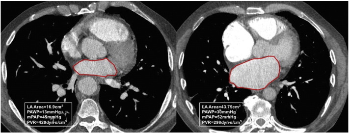
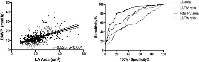
446 patients, of which 88 had left heart disease. Left atrial area was a strong predictor of elevated PAWP>15 mm Hg and PAWP>18 mm Hg, area under curve (AUC) 0.854, and AUC 0.873 respectively. Similar accuracy was also identified for MRI derived LA volume, AUC 0.852 and AUC 0.878 for PAWP > 15 and 18 mm Hg, respectively. Left atrial area of 26.8 cm and 30.0 cm were optimal specific thresholds for identification of PAWP > 15 and 18 mm Hg, had sensitivity of 60%/53% and specificity 89%/94%, respectively in a validation cohort.
CTPA and MRI derived left atrial size identifies left heart disease in suspected pulmonary hypertension with high specificity. The proposed diagnostic thresholds for elevated left atrial area on routine CTPA may be a useful to indicate the diagnosis of left heart disease in suspected pulmonary hypertension.
左心疾病(PH-LHD)导致的肺动脉高压患者与肺动脉高压患者具有重叠的临床特征,这使得诊断依赖于右心导管检查(RHC)。本研究旨在通过比较计算机断层肺动脉造影(CTPA)和磁共振成像(MRI)来研究 CT 肺动脉造影(CTPA)衍生的心肺结构指标,以诊断疑似肺动脉高压患者的左心疾病。
确定了疑似肺动脉高压且接受 CTPA、MRI 和 RHC 检查的患者。从 CTPA 和 MRI 记录心脏腔室和血管的测量值。在推导队列(n=235)中确定了检测升高的肺动脉楔压(PAWP)的各个测量值的诊断阈值。在验证队列(n=211)中测试了单独的 CT 和 MRI 衍生指标。
446 名患者中,88 名患有左心疾病。左心房面积是升高的 PAWP>15mmHg 和 PAWP>18mmHg 的强预测因子,曲线下面积(AUC)分别为 0.854 和 0.873。MRI 衍生的 LA 体积也具有相似的准确性,PAWP>15mmHg 和 18mmHg 的 AUC 分别为 0.852 和 0.878。LA 面积 26.8cm 和 30.0cm 分别是 PAWP>15mmHg 和 18mmHg 的最佳特异度截断值,在验证队列中的敏感性分别为 60%/53%,特异性分别为 89%/94%。
CTPA 和 MRI 衍生的左心房大小可以高度特异性地识别疑似肺动脉高压中的左心疾病。常规 CTPA 上升高的左心房面积的诊断阈值可能有助于提示疑似肺动脉高压中的左心疾病的诊断。