• 文献检索
  • 文档翻译
  • 深度研究
  • 学术资讯
  • Suppr Zotero 插件Zotero 插件
  • 邀请有礼
  • 套餐&价格
  • 历史记录
应用&插件
Suppr Zotero 插件Zotero 插件浏览器插件Mac 客户端Windows 客户端微信小程序
定价
高级版会员购买积分包购买API积分包
服务
文献检索文档翻译深度研究API 文档MCP 服务
关于我们
关于 Suppr公司介绍联系我们用户协议隐私条款
关注我们

Suppr 超能文献

核心技术专利:CN118964589B侵权必究
粤ICP备2023148730 号-1Suppr @ 2026

文献检索

告别复杂PubMed语法,用中文像聊天一样搜索,搜遍4000万医学文献。AI智能推荐,让科研检索更轻松。

立即免费搜索

文件翻译

保留排版,准确专业,支持PDF/Word/PPT等文件格式,支持 12+语言互译。

免费翻译文档

深度研究

AI帮你快速写综述,25分钟生成高质量综述,智能提取关键信息,辅助科研写作。

立即免费体验

使用眼前节光学相干断层扫描技术对角膜混浊进行客观量化。

Objective quantification of corneal haziness using anterior segment optical coherence tomography.

作者信息

Rose Jeyanth Suresh, Eldrina Juliet, Joshua Aarwin, Amalan S, Sebastian Tunny, Solomon Satheesh, Korah Sanita

机构信息

Department of Ophthalmology, Christian Medical College, Vellore, India.

Center for Stem Cell Research, Christian Medical College, Vellore, India.

出版信息

J Curr Ophthalmol. 2017 Sep 1;30(1):54-57. doi: 10.1016/j.joco.2017.08.001. eCollection 2018 Mar.

DOI:10.1016/j.joco.2017.08.001
PMID:29564409
原文链接:https://pmc.ncbi.nlm.nih.gov/articles/PMC5859339/
Abstract

PURPOSE

To quantify normal corneal transparency by anterior segment optical coherence tomography (AS-OCT) by measuring the average pixel intensity. To analyze the variation in the average pixel intensity in mild and severe grades of corneal opacities.

METHODS

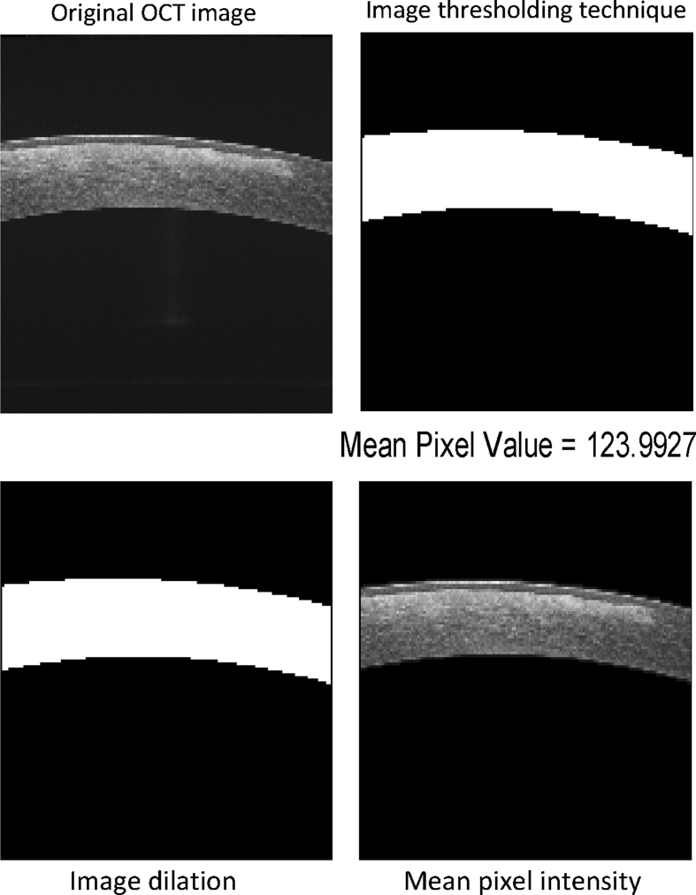
This is an observational, cross-sectional study of 38 eyes from 19 patients with mild or severe grades of corneal opacities greater than 3 mm and a normal contralateral cornea. AS-OCT was performed centered on the opacity with a 3 mm cruciate protocol. A similar image is taken of the contralateral clear cornea in the same quadrant. The average pixel intensity was calculated in a standardized manner using MATLAB software.

RESULT

The average pixel intensity of the normal cornea was 99.6 ± 10.9 [standard deviation (SD)]. The average pixel intensity of the mild and severe corneal opacities was 115.5 ± 9.1 and 141.1 ± 10.3, respectively. The differences were statistically significant.

CONCLUSIONS

AS-OCT images can be used to quantify corneal transparency. Average pixel intensity is a measure that varies significantly with varying corneal opacification.

摘要

目的

通过测量平均像素强度,利用眼前节光学相干断层扫描(AS-OCT)对正常角膜透明度进行量化。分析轻度和重度角膜混浊患者平均像素强度的变化情况。

方法

这是一项观察性横断面研究,纳入了19例角膜混浊程度为轻度或重度(混浊范围大于3mm)的患者的38只眼,对侧角膜正常。采用3mm十字形扫描方案,以混浊部位为中心进行AS-OCT检查。在同一象限对侧透明角膜拍摄类似图像。使用MATLAB软件以标准化方式计算平均像素强度。

结果

正常角膜的平均像素强度为99.6±10.9[标准差(SD)]。轻度和重度角膜混浊的平均像素强度分别为115.5±9.1和141.1±10.3。差异具有统计学意义。

结论

AS-OCT图像可用于量化角膜透明度。平均像素强度是一种随角膜混浊程度变化而有显著差异的测量指标。

https://cdn.ncbi.nlm.nih.gov/pmc/blobs/76b6/5859339/5880a903ba31/gr2.jpg
https://cdn.ncbi.nlm.nih.gov/pmc/blobs/76b6/5859339/71011f3ed285/gr1.jpg
https://cdn.ncbi.nlm.nih.gov/pmc/blobs/76b6/5859339/5880a903ba31/gr2.jpg
https://cdn.ncbi.nlm.nih.gov/pmc/blobs/76b6/5859339/71011f3ed285/gr1.jpg
https://cdn.ncbi.nlm.nih.gov/pmc/blobs/76b6/5859339/5880a903ba31/gr2.jpg

相似文献

1
Objective quantification of corneal haziness using anterior segment optical coherence tomography.使用眼前节光学相干断层扫描技术对角膜混浊进行客观量化。
J Curr Ophthalmol. 2017 Sep 1;30(1):54-57. doi: 10.1016/j.joco.2017.08.001. eCollection 2018 Mar.
2
High-speed optical coherence tomography of corneal opacities.角膜混浊的高速光学相干断层扫描
Ophthalmology. 2007 Jul;114(7):1278-85. doi: 10.1016/j.ophtha.2006.10.033. Epub 2007 Feb 20.
3
Anterior segment optical coherence tomography in congenital corneal opacities.先天性角膜混浊的眼前节光学相干断层扫描。
Ophthalmology. 2012 Dec;119(12):2450-7. doi: 10.1016/j.ophtha.2012.06.050. Epub 2012 Sep 5.
4
Corneal pachymetry mapping with high-speed optical coherence tomography.利用高速光学相干断层扫描技术进行角膜测厚成像
Ophthalmology. 2006 May;113(5):792-9.e2. doi: 10.1016/j.ophtha.2006.01.048.
5
Intraoperative changes in corneal structure during excimer laser phototherapeutic keratectomy (PTK) assessed by intraoperative optical coherence tomography.通过术中光学相干断层扫描评估准分子激光光治疗性角膜切削术(PTK)期间角膜结构的术中变化。
Graefes Arch Clin Exp Ophthalmol. 2018 Mar;256(3):575-581. doi: 10.1007/s00417-017-3867-7. Epub 2018 Jan 4.
6
Comparability of corneal thickness and opacity depth assessed by OCT and UBM.OCT 和 UBM 评估的角膜厚度和不透明度深度的可比性。
Graefes Arch Clin Exp Ophthalmol. 2021 Jul;259(7):1915-1923. doi: 10.1007/s00417-021-05161-9. Epub 2021 Mar 25.
7
Anterior chamber width measurement by high-speed optical coherence tomography.通过高速光学相干断层扫描测量前房宽度
Ophthalmology. 2005 Feb;112(2):238-44. doi: 10.1016/j.ophtha.2004.09.019.
8
Measurement of corneal and limbal epithelial thickness by anterior segment optical coherence tomography and in vivo confocal microscopy.通过眼前节光学相干断层扫描和活体共聚焦显微镜测量角膜和角膜缘上皮厚度。
BMC Ophthalmol. 2016 Sep 20;16(1):163. doi: 10.1186/s12886-016-0342-x.
9
New severity grading system for Fuchs endothelial corneal dystrophy using anterior segment optical coherence tomography.使用眼前节光学相干断层扫描技术的Fuchs内皮性角膜营养不良新严重程度分级系统
Acta Ophthalmol. 2021 Sep;99(6):e914-e921. doi: 10.1111/aos.14690. Epub 2020 Nov 30.
10
Establishing an objective biomarker for corneal cystinosis using a threshold-based Spectral domain optical coherence tomography imaging algorithm.利用基于阈值的谱域光学相干断层扫描成像算法建立角膜胱氨酸沉积症的客观生物标志物。
Acta Ophthalmol. 2021 Mar;99(2):e189-e195. doi: 10.1111/aos.14569. Epub 2020 Aug 24.

引用本文的文献

1
Characterization of Directional Reflectance in Corneal Tissue: A Comprehensive Optical Coherence Tomography Analysis.角膜组织中定向反射率的表征:全面光学相干断层扫描分析
Transl Vis Sci Technol. 2025 Apr 1;14(4):7. doi: 10.1167/tvst.14.4.7.
2
Corneal densitometry measurements comparison between anterior segment OCT and scheimpflug imaging.眼前节 OCT 和 Scheimpflug 成像的角膜密度测量比较。
Int Ophthalmol. 2024 Sep 25;44(1):392. doi: 10.1007/s10792-024-03309-0.
3
Loss of TRPM8 Exacerbate Herpes Simplex Keratitis Infection in Mice by Promoting the Infiltration of CD11b+ Ly6G+ Cells and Increasing the Viral Load in the Cornea.

本文引用的文献

1
Obtaining Thickness Maps of Corneal Layers Using the Optimal Algorithm for Intracorneal Layer Segmentation.使用角膜内层分割优化算法获取角膜各层厚度图。
Int J Biomed Imaging. 2016;2016:1420230. doi: 10.1155/2016/1420230. Epub 2016 May 9.
2
New Developments in Murine Imaging for Assessing Photoreceptor Degeneration In Vivo.用于体内评估光感受器退化的小鼠成像新技术
Adv Exp Med Biol. 2016;854:269-75. doi: 10.1007/978-3-319-17121-0_36.
3
Corneal structure and transparency.角膜结构与透明度。
TRPM8 的缺失通过促进 CD11b+Ly6G+细胞的浸润和增加角膜中的病毒载量,加重了小鼠单纯疱疹性角膜炎感染。
Invest Ophthalmol Vis Sci. 2023 Dec 1;64(15):24. doi: 10.1167/iovs.64.15.24.
4
Small incision lenticule extraction and femtosecond-assisted laser keratomileusis in patients with deep corneal opacity: case series.小切口透镜切除术和飞秒激光制瓣准分子原位角膜磨镶术治疗深层角膜混浊患者:病例系列
Int J Ophthalmol. 2023 Feb 18;16(2):301-308. doi: 10.18240/ijo.2023.02.19. eCollection 2023.
5
Case report: Utilization of neutral density filters for densitometry analysis of dense corneal opacities.病例报告:使用中性密度滤光片对致密角膜混浊进行密度测定分析
Am J Ophthalmol Case Rep. 2022 Jul 31;27:101672. doi: 10.1016/j.ajoc.2022.101672. eCollection 2022 Sep.
Prog Retin Eye Res. 2015 Nov;49:1-16. doi: 10.1016/j.preteyeres.2015.07.001. Epub 2015 Jul 2.
4
Human limbal biopsy-derived stromal stem cells prevent corneal scarring.人角膜缘活检来源的基质干细胞可预防角膜瘢痕形成。
Sci Transl Med. 2014 Dec 10;6(266):266ra172. doi: 10.1126/scitranslmed.3009644.
5
Fourier-domain optical coherence tomography imaging in keratoconus: a corneal structural classification.频域光学相干断层成像在圆锥角膜中的应用:角膜结构分类。
Ophthalmology. 2013 Dec;120(12):2403-2412. doi: 10.1016/j.ophtha.2013.05.027. Epub 2013 Aug 9.
6
Transplantation of human umbilical mesenchymal stem cells cures the corneal defects of mucopolysaccharidosis VII mice.人脐带间充质干细胞移植治愈黏多糖贮积症VII型小鼠的角膜缺损
Stem Cells. 2013 Oct;31(10):2116-26. doi: 10.1002/stem.1481.
7
Quantitative histology of contused lung tissue with comparison to computed tomography.挫伤肺组织的定量组织学及与计算机断层扫描的比较
Biomed Sci Instrum. 2008;44:225-30.
8
Collaborative Longitudinal Evaluation of Keratoconus (CLEK) Study: methods and findings to date.圆锥角膜协作纵向评估(CLEK)研究:方法及迄今的发现
Cont Lens Anterior Eye. 2007 Sep;30(4):223-32. doi: 10.1016/j.clae.2007.03.001. Epub 2007 May 3.
9
Trachoma in The Gambia.冈比亚的沙眼
Br J Ophthalmol. 1998 Aug;82(8):930-3. doi: 10.1136/bjo.82.8.930.
10
Micrometer-scale resolution imaging of the anterior eye in vivo with optical coherence tomography.利用光学相干断层扫描对活体眼前部进行微米级分辨率成像。
Arch Ophthalmol. 1994 Dec;112(12):1584-9. doi: 10.1001/archopht.1994.01090240090031.