Nishishita Rie, Morohashi Satoko, Seino Hiroko, Wu Yunyan, Yoshizawa Tadashi, Haga Toshihiro, Saito Kensuke, Hakamada Kenichi, Fukuda Shinsaku, Kijima Hiroshi
Department of Pathology and Bioscience, Hirosaki University Graduate School of Medicine, Hirosaki, Aomori 036-8562, Japan.
Department of Gastroenterology and Hematology, Hirosaki University Graduate School of Medicine, Hirosaki, Aomori 036-8562, Japan.
Oncol Lett. 2018 May;15(5):6195-6202. doi: 10.3892/ol.2018.8097. Epub 2018 Feb 21.
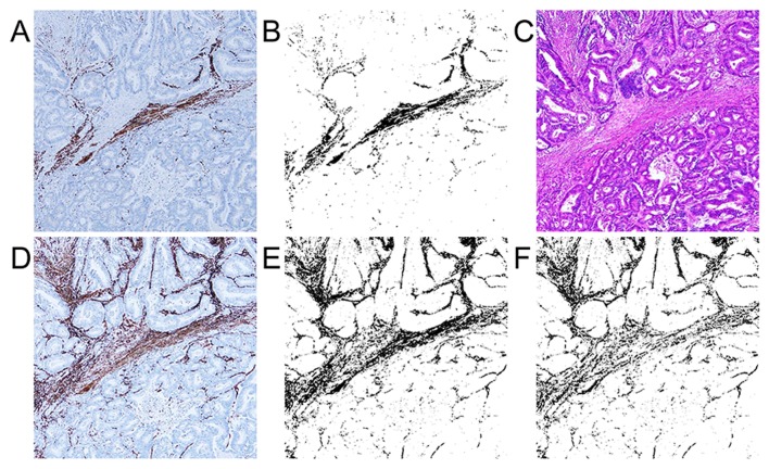
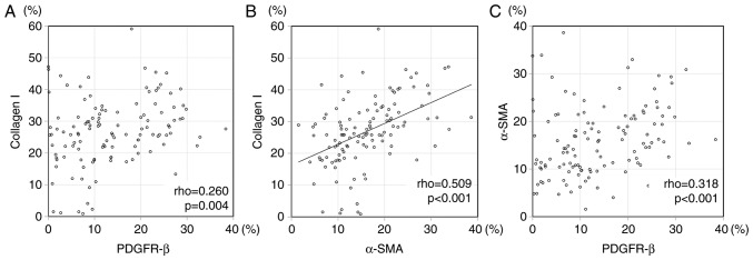
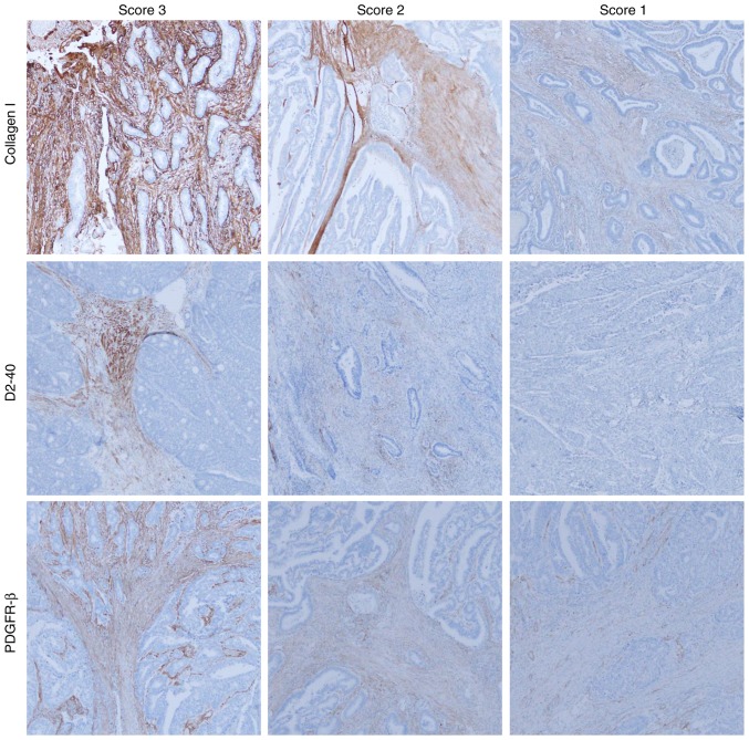
Colorectal cancer is one of the most common causes of mortality from cancer worldwide. Previous studies have demonstrated that cancer-associated fibroblasts (CAFs) promote neoangiogenesis and tumor growth for various tumors. The present study analyzed CAF markers, including α-smooth muscle actin (α-SMA), collagen I, platelet-derived growth factor receptor-β (PDGFR-β), and D2-40 (antibody recognizing podoplanin), and vessel markers, including cluster of differentiation (CD)31 and CD34, for 121 advanced colorectal cancer cases using a digital image analyzing technique. The association between CAF markers and vessel markers with clinicopathological factors was investigated. Furthermore, the association between CAF markers with each other, and their association with vessel markers was analyzed. Mean/median expression area of stromal and vessel markers in tumors were collagen I, 26.787%; D2-40, 1.372%; PDGFR-β, 11.646%; α-SMA-positive and desmin-negative myofibroblasts (α-SMA subtraction), 15.372%; CD31, 3.635%; and CD34, 2.226%. The expression area of α-SMA subtraction was significantly correlated with collagen I (P<0.001, correlation rho=0.509). High levels of α-SMA subtraction (P=0.002), collagen I (P=0.040), and PDGFR-β (P=0.040) expressions tended to be associated with high venous invasion. D2-40 did not correlate with other CAF and vessel markers. These results indicated that individual CAFs may have different expression patterns, and different strength effects for venous invasion in advanced colorectal cancer stroma.
结直肠癌是全球癌症死亡的最常见原因之一。先前的研究表明,癌症相关成纤维细胞(CAFs)可促进多种肿瘤的新生血管形成和肿瘤生长。本研究使用数字图像分析技术,分析了121例晚期结直肠癌病例的CAF标志物,包括α平滑肌肌动蛋白(α-SMA)、I型胶原蛋白、血小板衍生生长因子受体-β(PDGFR-β)和D2-40(识别血小板内皮细胞黏附分子的抗体),以及血管标志物,包括分化簇(CD)31和CD34。研究了CAF标志物和血管标志物与临床病理因素之间的关联。此外,还分析了CAF标志物之间的相互关联,以及它们与血管标志物的关联。肿瘤中基质和血管标志物的平均/中位表达面积分别为:I型胶原蛋白26.787%;D2-40 1.372%;PDGFR-β 11.646%;α-SMA阳性且结蛋白阴性的肌成纤维细胞(α-SMA减法)15.372%;CD31 3.635%;CD34 2.226%。α-SMA减法的表达面积与I型胶原蛋白显著相关(P<0.001,相关系数rho=0.509)。α-SMA减法(P=0.002)、I型胶原蛋白(P=0.040)和PDGFR-β(P=0.040)的高表达水平往往与高静脉侵犯相关。D2-40与其他CAF和血管标志物无相关性。这些结果表明,在晚期结直肠癌基质中,单个CAF可能具有不同的表达模式,对静脉侵犯的影响强度也不同。