Department of Integrative Biology and Physiology, University of Minnesota Medical School, Minneapolis, Minnesota, United States of America.
Department of Biomedical Engineering, University of Minnesota, Minneapolis, Minnesota, United States of America.
PLoS One. 2018 Apr 4;13(4):e0194909. doi: 10.1371/journal.pone.0194909. eCollection 2018.
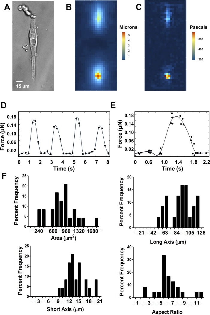
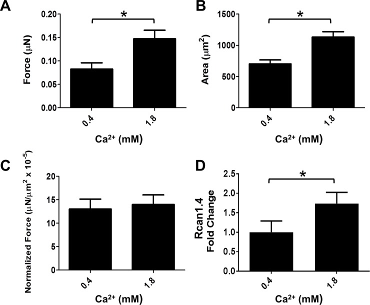
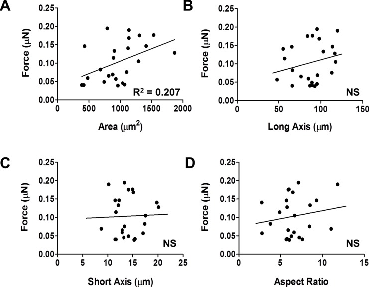
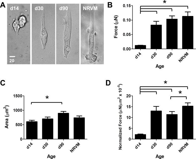
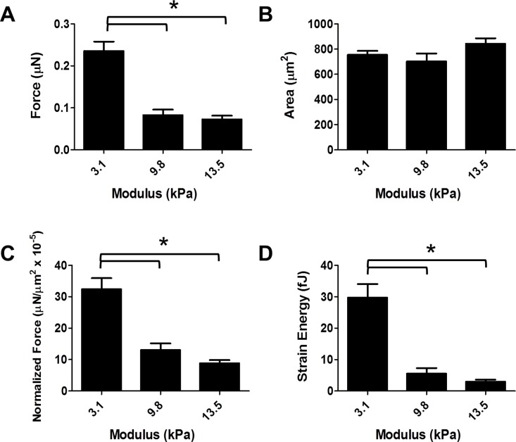
Recent advances have made it possible to readily derive cardiac myocytes from human induced pluripotent stem cells (hiPSC-CMs). HiPSC-CMs represent a valuable new experimental model for studying human cardiac muscle physiology and disease. Many laboratories have devoted substantial effort to examining the functional properties of isolated hiPSC-CMs, but to date, force production has not been adequately characterized. Here, we utilized traction force microscopy (TFM) with micro-patterning cell printing to investigate the maximum force production of isolated single hiPSC-CMs under varied culture and assay conditions. We examined the role of length of differentiation in culture and the effects of varied extracellular calcium concentration in the culture media on the maturation of hiPSC-CMs. Results show that hiPSC-CMs developing in culture for two weeks produced significantly less force than cells cultured from one to three months, with hiPSC-CMs cultured for three months resembling the cell morphology and function of neonatal rat ventricular myocytes in terms of size, dimensions, and force production. Furthermore, hiPSC-CMs cultured long term in conditions of physiologic calcium concentrations were larger and produced more force than hiPSC-CMs cultured in standard media with sub-physiological calcium. We also examined relationships between cell morphology, substrate stiffness and force production. Results showed a significant relationship between cell area and force. Implementing directed modifications of substrate stiffness, by varying stiffness from embryonic-like to adult myocardium-like, hiPSC-CMs produced maximal forces on substrates with a lower modulus and significantly less force when assayed on increasingly stiff adult myocardium-like substrates. Calculated strain energy measurements paralleled these findings. Collectively, these findings further establish single cell TFM as a valuable approach to illuminate the quantitative physiological maturation of force in hiPSC-CMs.
最近的进展使得从人类诱导多能干细胞(hiPSC-CMs)中轻易获得心肌细胞成为可能。hiPSC-CMs 代表了研究人类心肌生理学和疾病的一种有价值的新型实验模型。许多实验室都致力于研究分离的 hiPSC-CMs 的功能特性,但迄今为止,力的产生尚未得到充分描述。在这里,我们利用带有微图案细胞打印的牵引力显微镜(TFM)来研究在不同培养和检测条件下分离的单个 hiPSC-CMs 的最大力产生。我们研究了培养过程中分化时间的长度以及培养介质中不同细胞外钙浓度对 hiPSC-CMs 成熟的影响。结果表明,在培养中分化两周的 hiPSC-CMs 产生的力明显小于培养一到三个月的细胞,培养三个月的 hiPSC-CMs 在大小、尺寸和力产生方面与新生大鼠心室肌细胞的形态和功能相似。此外,长期在生理钙浓度条件下培养的 hiPSC-CMs 比在标准培养基中培养的 hiPSC-CMs 更大,产生的力也更多,而标准培养基中的钙浓度低于生理浓度。我们还研究了细胞形态、基质硬度和力产生之间的关系。结果表明细胞面积和力之间存在显著关系。通过改变基质硬度从胚胎样到成人心肌样,对基质硬度进行定向修饰,hiPSC-CMs 在低模量的基质上产生最大力,而在越来越硬的成人心肌样基质上检测时产生的力显著减小。计算出的应变能测量结果与这些发现一致。总的来说,这些发现进一步确立了单细胞 TFM 是一种有价值的方法,可以阐明 hiPSC-CMs 中力的定量生理成熟。