• 文献检索
  • 文档翻译
  • 深度研究
  • 学术资讯
  • Suppr Zotero 插件Zotero 插件
  • 邀请有礼
  • 套餐&价格
  • 历史记录
应用&插件
Suppr Zotero 插件Zotero 插件浏览器插件Mac 客户端Windows 客户端微信小程序
定价
高级版会员购买积分包购买API积分包
服务
文献检索文档翻译深度研究API 文档MCP 服务
关于我们
关于 Suppr公司介绍联系我们用户协议隐私条款
关注我们

Suppr 超能文献

核心技术专利:CN118964589B侵权必究
粤ICP备2023148730 号-1Suppr @ 2026

文献检索

告别复杂PubMed语法,用中文像聊天一样搜索,搜遍4000万医学文献。AI智能推荐,让科研检索更轻松。

立即免费搜索

文件翻译

保留排版,准确专业,支持PDF/Word/PPT等文件格式,支持 12+语言互译。

免费翻译文档

深度研究

AI帮你快速写综述,25分钟生成高质量综述,智能提取关键信息,辅助科研写作。

立即免费体验

使用3T高分辨率定量磁化率成像结合黑质直方图分析诊断早期特发性帕金森病

Diagnosis of Early-Stage Idiopathic Parkinson's Disease Using High-Resolution Quantitative Susceptibility Mapping Combined with Histogram Analysis in the Substantia Nigra at 3 T.

作者信息

Kim Eung Yeop, Sung Young Hee, Shin Hyeong Geol, Noh Young, Nam Yoonho, Lee Jongho

机构信息

Department of Radiology, Gachon University Gil Medical Center, Incheon, Korea.

Department of Neurology, Gachon University Gil Medical Center, Incheon, Korea.

出版信息

J Clin Neurol. 2018 Jan;14(1):90-97. doi: 10.3988/jcn.2018.14.1.90.

DOI:10.3988/jcn.2018.14.1.90
PMID:29629545
原文链接:https://pmc.ncbi.nlm.nih.gov/articles/PMC5765262/
Abstract

BACKGROUND AND PURPOSE

To test whether nigrosome-1 imaging using high-resolution quantitative susceptibility mapping (QSM) combined with histogram analysis can improve the diagnostic accuracy in early-stage idiopathic Parkinson's disease (IPD) patients.

METHODS

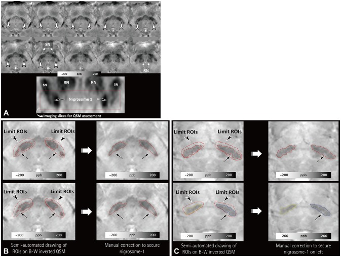
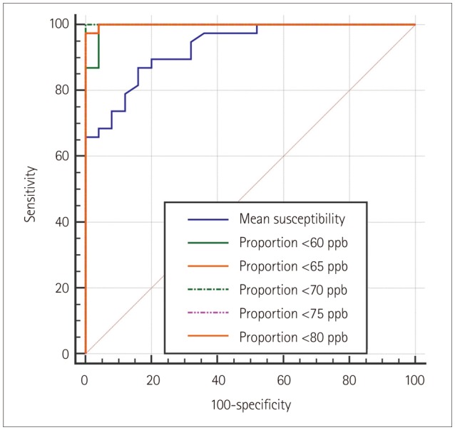
Three-dimensional multiecho gradient-recalled echo images (0.5×0.5×1.0 mm³) were obtained at 3 T for QSM in 38 patients with IPD and 25 healthy subjects. To segment the substantia nigra (SN), regions of interest (ROIs) were semiautomatically drawn at the location below the red nucleus, and the normal-appearing nigrosome-1 was determined by manual correction. QSM histograms were obtained within the ROI. The segmented SN regions on the right and left that had higher mean susceptibility values and fewer voxels with susceptibility values lower than 60, 65, 70, 75, and 80 ppb were chosen for comparisons between the IPD patients and healthy subjects. These results were compared with those of the visual assessments of nigrosome-1 in susceptibility map-weighted imaging (SMWI) by analyzing receiver operating characteristics curves.

RESULTS

The proportion of voxels with susceptibility values lower than 70 ppb showed the best diagnostic performance, with its value differing significantly between the IPD patients (median=0, interquartile range=0-0.23) and healthy subjects (median=10.67, interquartile range=5.98-21.57) (p<0.0001). The number of voxels with susceptibility values lower than 60, 65, 70, 75, and 80 ppb showed worse diagnostic performances but were still significantly better than that of the mean susceptibility value (p=0.0249, 0.0192, 0.0183, 0.0191, and 0.0186, respectively), which also differed significantly between the two groups: 125.81±16.27 ppb (mean±standard deviation) in IPD versus 98.41±11.70 ppb in healthy subjects (p<0.0001). Additionally, using the proportion of voxels with susceptibility values lower than 70 ppb provided significantly better diagnostic performance than did visual assessments of SMWI (p=0.0143).

CONCLUSIONS

High-spatial-resolution QSM combined with histogram analysis at 3 T can improve the diagnostic accuracy of early-stage IPD.

摘要

背景与目的

测试使用高分辨率定量磁化率成像(QSM)结合直方图分析的黑质1成像是否能提高早期特发性帕金森病(IPD)患者的诊断准确性。

方法

在3T条件下,对38例IPD患者和25名健康受试者进行三维多回波梯度回波成像(0.5×0.5×1.0mm³)以获取QSM。为分割黑质(SN),在红核下方位置半自动绘制感兴趣区域(ROI),并通过手动校正确定外观正常的黑质1。在ROI内获取QSM直方图。选择左右两侧分割的SN区域,其平均磁化率值较高且磁化率值低于60、65、70、75和80ppb的体素较少,用于IPD患者与健康受试者之间的比较。通过分析受试者工作特征曲线,将这些结果与在磁化率图加权成像(SMWI)中对黑质1的视觉评估结果进行比较。

结果

磁化率值低于70ppb的体素比例显示出最佳诊断性能,其值在IPD患者(中位数=0,四分位间距=0 - 0.23)和健康受试者(中位数=10.67,四分位间距=5.98 - 21.57)之间存在显著差异(p<0.0001)。磁化率值低于60、65、70、75和80ppb的体素数量显示出较差的诊断性能,但仍显著优于平均磁化率值(p分别为0.0249、0.0192、0.0183、0.0191和0.0186),两组之间也存在显著差异:IPD患者为125.81±16.27ppb(均值±标准差),健康受试者为98.41±11.70ppb(p<0.0001)。此外,使用磁化率值低于70ppb的体素比例提供的诊断性能明显优于SMWI的视觉评估(p=0.0143)。

结论

3T条件下的高空间分辨率QSM结合直方图分析可提高早期IPD的诊断准确性。

https://cdn.ncbi.nlm.nih.gov/pmc/blobs/97fd/5765262/848ffcaefe5c/jcn-14-90-g003.jpg
https://cdn.ncbi.nlm.nih.gov/pmc/blobs/97fd/5765262/92ef212c89c2/jcn-14-90-g001.jpg
https://cdn.ncbi.nlm.nih.gov/pmc/blobs/97fd/5765262/2940b1aff2bb/jcn-14-90-g002.jpg
https://cdn.ncbi.nlm.nih.gov/pmc/blobs/97fd/5765262/848ffcaefe5c/jcn-14-90-g003.jpg
https://cdn.ncbi.nlm.nih.gov/pmc/blobs/97fd/5765262/92ef212c89c2/jcn-14-90-g001.jpg
https://cdn.ncbi.nlm.nih.gov/pmc/blobs/97fd/5765262/2940b1aff2bb/jcn-14-90-g002.jpg
https://cdn.ncbi.nlm.nih.gov/pmc/blobs/97fd/5765262/848ffcaefe5c/jcn-14-90-g003.jpg

相似文献

1
Diagnosis of Early-Stage Idiopathic Parkinson's Disease Using High-Resolution Quantitative Susceptibility Mapping Combined with Histogram Analysis in the Substantia Nigra at 3 T.使用3T高分辨率定量磁化率成像结合黑质直方图分析诊断早期特发性帕金森病
J Clin Neurol. 2018 Jan;14(1):90-97. doi: 10.3988/jcn.2018.14.1.90.
2
A preliminary attempt to visualize nigrosome 1 in the substantia nigra for Parkinson's disease at 3T: An efficient susceptibility map-weighted imaging (SMWI) with quantitative susceptibility mapping using deep neural network (QSMnet).初步尝试在 3T 下可视化帕金森病黑质 1:使用深度神经网络(QSMnet)进行定量磁化率映射的有效磁化率图加权成像(SMWI)。
Med Phys. 2020 Mar;47(3):1151-1160. doi: 10.1002/mp.13999. Epub 2020 Jan 30.
3
Radiomic Features of the Nigrosome-1 Region of the Substantia Nigra: Using Quantitative Susceptibility Mapping to Assist the Diagnosis of Idiopathic Parkinson's Disease.黑质黑质小体1区的放射组学特征:利用定量磁化率成像辅助特发性帕金森病的诊断。
Front Aging Neurosci. 2019 Jul 16;11:167. doi: 10.3389/fnagi.2019.00167. eCollection 2019.
4
Imaging of nigrosome 1 in substantia nigra at 3T using multiecho susceptibility map-weighted imaging (SMWI).使用多回波磁化率图加权成像(SMWI)在 3T 下对黑质内的 nigrosome 1 进行成像。
J Magn Reson Imaging. 2017 Aug;46(2):528-536. doi: 10.1002/jmri.25553. Epub 2016 Nov 11.
5
Early-stage Parkinson's disease: Abnormal nigrosome 1 and 2 revealed by a voxelwise analysis of neuromelanin-sensitive MRI.早期帕金森病:基于神经黑色素敏感 MRI 的体素分析显示的黑质 1 区和 2 区异常。
Hum Brain Mapp. 2021 Jun 15;42(9):2823-2832. doi: 10.1002/hbm.25406. Epub 2021 Mar 10.
6
Imaging the Nigrosome 1 in the substantia nigra using susceptibility weighted imaging and quantitative susceptibility mapping: An application to Parkinson's disease.应用磁敏感加权成像和定量磁敏感图对黑质 1 进行成像:在帕金森病中的应用。
Neuroimage Clin. 2020;25:102103. doi: 10.1016/j.nicl.2019.102103. Epub 2019 Nov 20.
7
The applied value in brain gray matter nuclei of patients with early-stage Parkinson's disease : a study based on multiple magnetic resonance imaging techniques.早期帕金森病患者脑灰质核中的应用价值:基于多种磁共振成像技术的研究。
Head Face Med. 2023 Jun 29;19(1):25. doi: 10.1186/s13005-023-00371-4.
8
Differential involvement of nigral subregions in idiopathic parkinson's disease.黑质亚区域在特发性帕金森病中的不同受累情况。
Hum Brain Mapp. 2018 Jan;39(1):542-553. doi: 10.1002/hbm.23863. Epub 2017 Oct 24.
9
Diagnostic performance of a high-spatial-resolution voxelwise analysis of neuromelanin-sensitive imaging in early-stage idiopathic Parkinson's disease.高空间分辨率体素分析神经黑色素敏感成像在早期特发性帕金森病中的诊断性能。
BMC Med Imaging. 2023 May 18;23(1):64. doi: 10.1186/s12880-023-01018-1.
10
Classification of Parkinson's disease by deep learning on midbrain MRI.基于中脑磁共振成像的深度学习对帕金森病的分类
Front Aging Neurosci. 2024 Aug 20;16:1425095. doi: 10.3389/fnagi.2024.1425095. eCollection 2024.

引用本文的文献

1
Automated segmentation of midbrain nuclei using deep learning and multisequence MRI: A longitudinal study on iron accumulation with age.使用深度学习和多序列磁共振成像对中脑核进行自动分割:关于铁蓄积与年龄关系的纵向研究。
Imaging Neurosci (Camb). 2024 Oct 7;2. doi: 10.1162/imag_a_00304. eCollection 2024.
2
Susceptibility-Weighted Imaging (SWI): Technical Aspects and Applications in Brain MRI for Neurodegenerative Disorders.磁敏感加权成像(SWI):脑MRI在神经退行性疾病中的技术要点与应用
Bioengineering (Basel). 2025 Apr 29;12(5):473. doi: 10.3390/bioengineering12050473.
3
High-resolution mapping of substantia nigra in Parkinson's disease using 7 tesla magnetic resonance imaging.

本文引用的文献

1
Can loss of the swallow tail sign help distinguish between Parkinson Disease and the Parkinson-Plus syndromes?燕尾征消失能否有助于鉴别帕金森病和帕金森叠加综合征?
Clin Imaging. 2017 Jul-Aug;44:66-69. doi: 10.1016/j.clinimag.2017.04.005. Epub 2017 Apr 20.
2
Using 'swallow-tail' sign and putaminal hypointensity as biomarkers to distinguish multiple system atrophy from idiopathic Parkinson's disease: A susceptibility-weighted imaging study.使用“燕尾征”和壳核低信号作为生物标志物区分多系统萎缩与特发性帕金森病:一项磁敏感加权成像研究
Eur Radiol. 2017 Aug;27(8):3174-3180. doi: 10.1007/s00330-017-4743-x. Epub 2017 Jan 19.
3
使用7特斯拉磁共振成像对帕金森病黑质进行高分辨率图谱绘制。
NPJ Parkinsons Dis. 2025 May 6;11(1):113. doi: 10.1038/s41531-025-00972-7.
4
Susceptibility map-weighted MRI can distinguish tremor-dominant Parkinson's disease from essential tremor.磁化率图加权磁共振成像能够区分震颤为主型帕金森病与特发性震颤。
Sci Rep. 2025 Jan 4;15(1):823. doi: 10.1038/s41598-024-81089-4.
5
Deep Learning-Based Algorithm for Automatic Quantification of Nigrosome-1 and Parkinsonism Classification Using Susceptibility Map-Weighted MRI.基于深度学习的算法,利用磁化率图加权磁共振成像自动定量黑质1和进行帕金森症分类
AJNR Am J Neuroradiol. 2025 May 2;46(5):999-1006. doi: 10.3174/ajnr.A8585.
6
Classification of Parkinson's disease by deep learning on midbrain MRI.基于中脑磁共振成像的深度学习对帕金森病的分类
Front Aging Neurosci. 2024 Aug 20;16:1425095. doi: 10.3389/fnagi.2024.1425095. eCollection 2024.
7
Neuroimaging and fluid biomarkers in Parkinson's disease in an era of targeted interventions.神经影像学和液体生物标志物在靶向干预时代的帕金森病。
Nat Commun. 2024 Jul 5;15(1):5661. doi: 10.1038/s41467-024-49949-9.
8
Iron deposition in subcortical nuclei of Parkinson's disease: A meta-analysis of quantitative iron-sensitive magnetic resonance imaging studies.帕金森病皮层下核团铁沉积:基于定量铁敏感磁共振成像研究的荟萃分析
Chin Med J (Engl). 2025 Mar 20;138(6):678-692. doi: 10.1097/CM9.0000000000003167. Epub 2024 May 28.
9
Loss of the "swallow tail sign" on susceptibility-weighted imaging in the diagnosis of dementia with Lewy bodies: a systematic review and meta-analysis.路易体痴呆症患者的磁敏感加权成像上“燕尾征”缺失的诊断价值:系统评价和荟萃分析。
J Neurol. 2024 Jul;271(7):3754-3763. doi: 10.1007/s00415-024-12381-6. Epub 2024 May 27.
10
So You Want to Image Myelin Using MRI: Magnetic Susceptibility Source Separation for Myelin Imaging.那么,你想用 MRI 对髓鞘成像:用于髓鞘成像的磁共振磁化率源分离。
Magn Reson Med Sci. 2024 Jul 1;23(3):291-306. doi: 10.2463/mrms.rev.2024-0001. Epub 2024 Apr 20.
9.4 T MR microscopy of the substantia nigra with pathological validation in controls and disease.
9.4 对黑质进行9.4T磁共振显微镜检查,并在对照组和疾病组中进行病理验证。
Neuroimage Clin. 2016 Nov 17;13:154-163. doi: 10.1016/j.nicl.2016.11.015. eCollection 2017.
4
Imaging of nigrosome 1 in substantia nigra at 3T using multiecho susceptibility map-weighted imaging (SMWI).使用多回波磁化率图加权成像(SMWI)在 3T 下对黑质内的 nigrosome 1 进行成像。
J Magn Reson Imaging. 2017 Aug;46(2):528-536. doi: 10.1002/jmri.25553. Epub 2016 Nov 11.
5
The whole-brain pattern of magnetic susceptibility perturbations in Parkinson's disease.帕金森病的全脑磁化率扰动模式。
Brain. 2017 Jan;140(1):118-131. doi: 10.1093/brain/aww278. Epub 2016 Nov 11.
6
Quantitative Susceptibility Mapping in Parkinson's Disease.帕金森病中的定量磁化率成像
PLoS One. 2016 Sep 6;11(9):e0162460. doi: 10.1371/journal.pone.0162460. eCollection 2016.
7
Loss of Nigral Hyperintensity on 3 Tesla MRI of Parkinsonism: Comparison With (123) I-FP-CIT SPECT.帕金森病3特斯拉磁共振成像黑质高信号缺失:与(123)I-FP-CIT单光子发射计算机断层扫描的比较
Mov Disord. 2016 May;31(5):684-92. doi: 10.1002/mds.26584. Epub 2016 Mar 17.
8
Nigral involvement in atypical parkinsonisms: evidence from a pilot study with ultra-high field MRI.
J Neural Transm (Vienna). 2016 May;123(5):509-13. doi: 10.1007/s00702-016-1529-2. Epub 2016 Mar 4.
9
Regionally progressive accumulation of iron in Parkinson's disease as measured by quantitative susceptibility mapping.通过定量磁化率成像测量帕金森病中铁的区域渐进性积累。
NMR Biomed. 2017 Apr;30(4). doi: 10.1002/nbm.3489. Epub 2016 Feb 8.
10
Lateral Asymmetry and Spatial Difference of Iron Deposition in the Substantia Nigra of Patients with Parkinson Disease Measured with Quantitative Susceptibility Mapping.帕金森病患者黑质铁沉积的侧方不对称性及空间差异的定量磁化率成像测量
AJNR Am J Neuroradiol. 2016 May;37(5):782-8. doi: 10.3174/ajnr.A4645. Epub 2016 Jan 28.