• 文献检索
  • 文档翻译
  • 深度研究
  • 学术资讯
  • Suppr Zotero 插件Zotero 插件
  • 邀请有礼
  • 套餐&价格
  • 历史记录
应用&插件
Suppr Zotero 插件Zotero 插件浏览器插件Mac 客户端Windows 客户端微信小程序
定价
高级版会员购买积分包购买API积分包
服务
文献检索文档翻译深度研究API 文档MCP 服务
关于我们
关于 Suppr公司介绍联系我们用户协议隐私条款
关注我们

Suppr 超能文献

核心技术专利:CN118964589B侵权必究
粤ICP备2023148730 号-1Suppr @ 2026

文献检索

告别复杂PubMed语法,用中文像聊天一样搜索,搜遍4000万医学文献。AI智能推荐,让科研检索更轻松。

立即免费搜索

文件翻译

保留排版,准确专业,支持PDF/Word/PPT等文件格式,支持 12+语言互译。

免费翻译文档

深度研究

AI帮你快速写综述,25分钟生成高质量综述,智能提取关键信息,辅助科研写作。

立即免费体验

黑质黑质小体1区的放射组学特征:利用定量磁化率成像辅助特发性帕金森病的诊断。

Radiomic Features of the Nigrosome-1 Region of the Substantia Nigra: Using Quantitative Susceptibility Mapping to Assist the Diagnosis of Idiopathic Parkinson's Disease.

作者信息

Cheng Zenghui, Zhang Jiping, He Naying, Li Yan, Wen Yaofeng, Xu Hongmin, Tang Rongbiao, Jin Zhijia, Haacke E Mark, Yan Fuhua, Qian Dahong

机构信息

Department of Radiology, Ruijin Hospital, Shanghai Jiao Tong University School of Medicine, Shanghai, China.

School of Biomedical Engineering, Shanghai Jiao Tong University, Shanghai, China.

出版信息

Front Aging Neurosci. 2019 Jul 16;11:167. doi: 10.3389/fnagi.2019.00167. eCollection 2019.

DOI:10.3389/fnagi.2019.00167
PMID:31379555
原文链接:https://pmc.ncbi.nlm.nih.gov/articles/PMC6648885/
Abstract

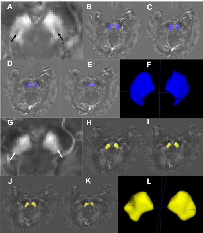
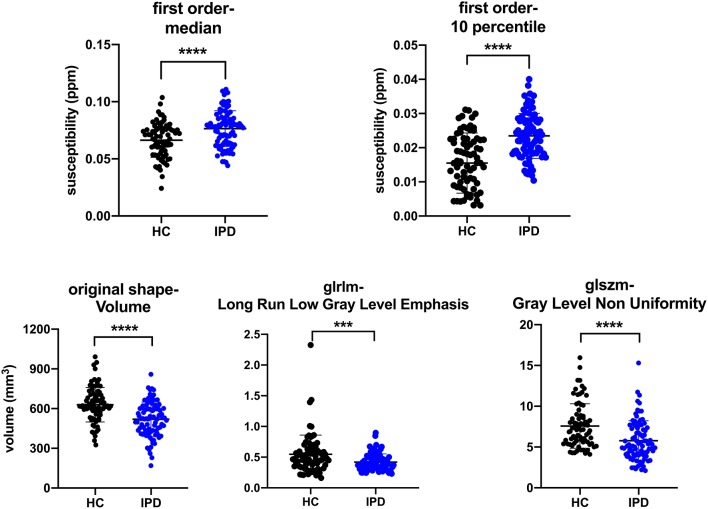
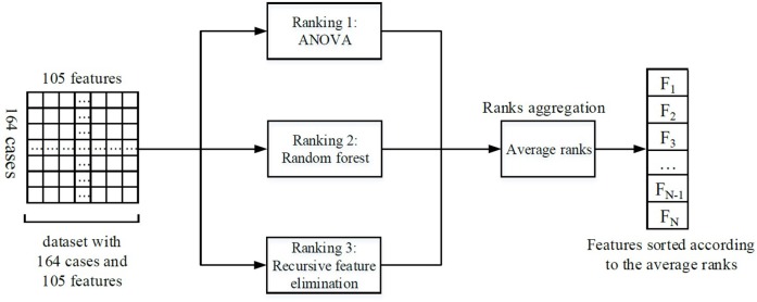
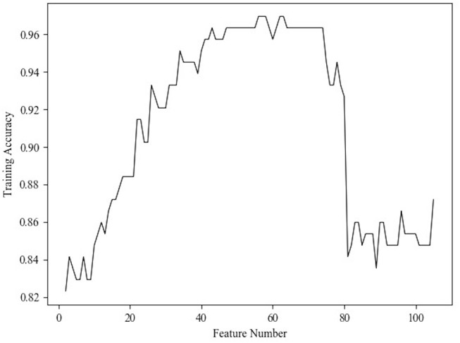
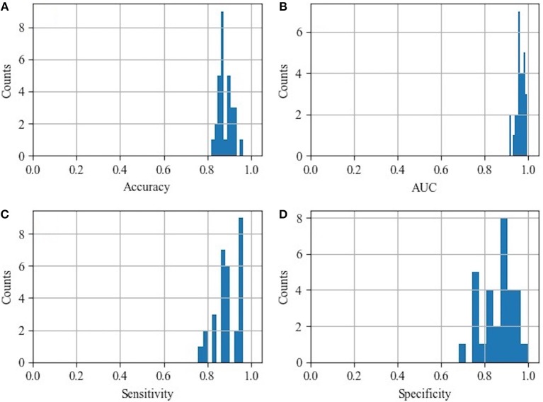
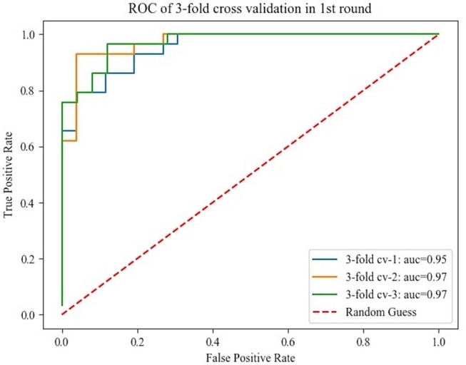
The loss of nigrosome-1, which is also referred to as the swallow tail sign (STS) in T2-weighted iron-sensitive magnetic resonance imaging (MRI), has recently emerged as a new biomarker for idiopathic Parkinson's disease (IPD). However, consistent recognition of the STS is difficult due to individual variations and different imaging parameters. Radiomics might have the potential to overcome these shortcomings. Therefore, we chose to explore whether radiomic features of nigrosome-1 of substantia nigra (SN) based on quantitative susceptibility mapping (QSM) could help to differentiate IPD patients from healthy controls (HCs). Three-dimensional multi-echo gradient-recalled echo images (0.86 × 0.86 × 1.00 mm) were obtained at 3.0-T MRI for QSM in 87 IPD patients and 77 HCs. Regions of interest (ROIs) of the SN below the red nucleus were manually drawn on both sides, and subsequently, volumes of interest (VOIs) were segmented (these ROIs included four 1-mm slices). Then, 105 radiomic features (including 18 first-order features, 13 shape features, and 74 texture features) of bilateral VOIs in the two groups were extracted. Forty features were selected according to the ensemble feature selection method, which combined analysis of variance, random forest, and recursive feature elimination. The selected features were further utilized to distinguish IPD patients from HC using the SVM classifier with 10 rounds of 3-fold cross-validation. Finally, the representative features were analyzed using an unpaired -test with Bonferroni correction and correlated with the UPDRS-III scores. The classification results from SVM were as follows: area under curve (AUC): 0.96 ± 0.02; accuracy: 0.88 ± 0.03; sensitivity: 0.89 ± 0.06; and specificity: 0.87 ± 0.07. Five representative features were selected to show their detailed difference between IPD patients and HCs: 10th percentile and median in IPD patients were higher than those in HCs (all < 0.00125), while Gray Level Run Length Matrix (GLRLM)-Long Run Low Gray Level Emphasis, Gray Level Size Zone Matrix (GLSZM)-Gray Level Non-Uniformity, and volume (all < 0.00125) in IPD patients were lower than those in HCs. The 10th percentile was positively correlated with UPDRS-III score ( = 0.35, = 0.001). Radiomic features of the nigrosome-1 region of SN based on QSM could be useful in the diagnosis of IPD and could serve as a surrogate marker for the STS.

摘要

在T2加权铁敏感磁共振成像(MRI)中,黑质小体-1的缺失,也被称为燕尾征(STS),最近已成为特发性帕金森病(IPD)的一种新生物标志物。然而,由于个体差异和不同的成像参数,对STS的一致识别较为困难。放射组学可能有潜力克服这些缺点。因此,我们选择探讨基于定量磁化率映射(QSM)的黑质(SN)黑质小体-1的放射组学特征是否有助于区分IPD患者和健康对照(HCs)。在3.0-T MRI上,对87例IPD患者和77例HCs进行QSM检查,获取三维多回波梯度回波图像(0.86×0.86×1.00 mm)。在双侧手动绘制红核下方SN的感兴趣区域(ROIs),随后分割感兴趣体积(VOIs)(这些ROIs包括四个1-mm切片)。然后,提取两组双侧VOIs的105个放射组学特征(包括18个一阶特征、13个形状特征和74个纹理特征)。根据综合特征选择方法选择40个特征,该方法结合了方差分析、随机森林和递归特征消除。使用支持向量机(SVM)分类器,通过10轮3折交叉验证,进一步利用所选特征区分IPD患者和HC。最后,使用Bonferroni校正的非配对t检验分析代表性特征,并与统一帕金森病评定量表第三部分(UPDRS-III)评分相关。SVM的分类结果如下:曲线下面积(AUC):0.96±0.02;准确率:0.88±0.03;灵敏度:0.89±0.06;特异性:0.87±0.07。选择五个代表性特征以显示IPD患者和HCs之间的详细差异:IPD患者的第10百分位数和中位数高于HCs(均P<0.00125),而IPD患者的灰度游程长度矩阵(GLRLM)-长游程低灰度强调、灰度大小区域矩阵(GLSZM)-灰度不均匀性和体积(均P<0.00125)低于HCs。第10百分位数与UPDRS-III评分呈正相关(r=0.35,P=0.001)。基于QSM的SN黑质小体-1区域的放射组学特征可用于IPD的诊断,并可作为STS的替代标志物。

https://cdn.ncbi.nlm.nih.gov/pmc/blobs/1ab4/6648885/bd4d05899698/fnagi-11-00167-g0006.jpg
https://cdn.ncbi.nlm.nih.gov/pmc/blobs/1ab4/6648885/4c0ac5baaede/fnagi-11-00167-g0001.jpg
https://cdn.ncbi.nlm.nih.gov/pmc/blobs/1ab4/6648885/3498daf491b1/fnagi-11-00167-g0002.jpg
https://cdn.ncbi.nlm.nih.gov/pmc/blobs/1ab4/6648885/6f762349a682/fnagi-11-00167-g0003.jpg
https://cdn.ncbi.nlm.nih.gov/pmc/blobs/1ab4/6648885/04186e60019c/fnagi-11-00167-g0004.jpg
https://cdn.ncbi.nlm.nih.gov/pmc/blobs/1ab4/6648885/51ee749caad8/fnagi-11-00167-g0005.jpg
https://cdn.ncbi.nlm.nih.gov/pmc/blobs/1ab4/6648885/bd4d05899698/fnagi-11-00167-g0006.jpg

相似文献

1
Radiomic Features of the Nigrosome-1 Region of the Substantia Nigra: Using Quantitative Susceptibility Mapping to Assist the Diagnosis of Idiopathic Parkinson's Disease.黑质黑质小体1区的放射组学特征:利用定量磁化率成像辅助特发性帕金森病的诊断。
Front Aging Neurosci. 2019 Jul 16;11:167. doi: 10.3389/fnagi.2019.00167. eCollection 2019.
2
Imaging the Nigrosome 1 in the substantia nigra using susceptibility weighted imaging and quantitative susceptibility mapping: An application to Parkinson's disease.应用磁敏感加权成像和定量磁敏感图对黑质 1 进行成像:在帕金森病中的应用。
Neuroimage Clin. 2020;25:102103. doi: 10.1016/j.nicl.2019.102103. Epub 2019 Nov 20.
3
Diagnosis of Early-Stage Idiopathic Parkinson's Disease Using High-Resolution Quantitative Susceptibility Mapping Combined with Histogram Analysis in the Substantia Nigra at 3 T.使用3T高分辨率定量磁化率成像结合黑质直方图分析诊断早期特发性帕金森病
J Clin Neurol. 2018 Jan;14(1):90-97. doi: 10.3988/jcn.2018.14.1.90.
4
A preliminary attempt to visualize nigrosome 1 in the substantia nigra for Parkinson's disease at 3T: An efficient susceptibility map-weighted imaging (SMWI) with quantitative susceptibility mapping using deep neural network (QSMnet).初步尝试在 3T 下可视化帕金森病黑质 1:使用深度神经网络(QSMnet)进行定量磁化率映射的有效磁化率图加权成像(SMWI)。
Med Phys. 2020 Mar;47(3):1151-1160. doi: 10.1002/mp.13999. Epub 2020 Jan 30.
5
The applied value in brain gray matter nuclei of patients with early-stage Parkinson's disease : a study based on multiple magnetic resonance imaging techniques.早期帕金森病患者脑灰质核中的应用价值:基于多种磁共振成像技术的研究。
Head Face Med. 2023 Jun 29;19(1):25. doi: 10.1186/s13005-023-00371-4.
6
MRI-Based Radiomics of Basal Nuclei in Differentiating Idiopathic Parkinson's Disease From Parkinsonian Variants of Multiple System Atrophy: A Susceptibility-Weighted Imaging Study.基于磁共振成像的基底核放射组学在鉴别特发性帕金森病与多系统萎缩帕金森综合征中的应用:一项磁敏感加权成像研究
Front Aging Neurosci. 2020 Nov 12;12:587250. doi: 10.3389/fnagi.2020.587250. eCollection 2020.
7
Diagnosing Parkinson's disease by combining neuromelanin and iron imaging features using an automated midbrain template approach.使用自动中脑模板方法结合神经黑色素和铁成像特征诊断帕金森病。
Neuroimage. 2023 Feb 1;266:119814. doi: 10.1016/j.neuroimage.2022.119814. Epub 2022 Dec 14.
8
Imaging iron and neuromelanin simultaneously using a single 3D gradient echo magnetization transfer sequence: Combining neuromelanin, iron and the nigrosome-1 sign as complementary imaging biomarkers in early stage Parkinson's disease.使用单个 3D 梯度回波磁化传递序列同时对铁和神经黑色素进行成像:将神经黑色素、铁和黑质-1 征作为早期帕金森病的互补成像生物标志物进行联合分析。
Neuroimage. 2021 Apr 15;230:117810. doi: 10.1016/j.neuroimage.2021.117810. Epub 2021 Jan 30.
9
Imaging of nigrosome 1 in substantia nigra at 3T using multiecho susceptibility map-weighted imaging (SMWI).使用多回波磁化率图加权成像(SMWI)在 3T 下对黑质内的 nigrosome 1 进行成像。
J Magn Reson Imaging. 2017 Aug;46(2):528-536. doi: 10.1002/jmri.25553. Epub 2016 Nov 11.
10
Quantifying iron deposition within the substantia nigra of Parkinson's disease by quantitative susceptibility mapping.采用定量磁化率映射技术对帕金森病患者黑质内铁沉积进行定量分析。
J Neurol Sci. 2018 Mar 15;386:46-52. doi: 10.1016/j.jns.2018.01.008. Epub 2018 Jan 12.

引用本文的文献

1
Dorsal hyperintensity and iron deposition patterns in the substantia nigra of Parkinson's disease, idiopathic REM sleep behavior disorder, and Parkinson-plus syndromes at 7T MRI: a prospective diagnostic study.7T磁共振成像下帕金森病、特发性快速眼动睡眠行为障碍及帕金森叠加综合征患者黑质的背侧高信号和铁沉积模式:一项前瞻性诊断研究
Transl Neurodegener. 2025 Jul 4;14(1):35. doi: 10.1186/s40035-025-00495-4.
2
Susceptibility-Weighted Imaging (SWI): Technical Aspects and Applications in Brain MRI for Neurodegenerative Disorders.磁敏感加权成像(SWI):脑MRI在神经退行性疾病中的技术要点与应用
Bioengineering (Basel). 2025 Apr 29;12(5):473. doi: 10.3390/bioengineering12050473.
3

本文引用的文献

1
Robust Ensemble Classification Methodology for I123-Ioflupane SPECT Images and Multiple Heterogeneous Biomarkers in the Diagnosis of Parkinson's Disease.用于帕金森病诊断的¹²³I-碘氟烷单光子发射计算机断层扫描(SPECT)图像和多种异质生物标志物的稳健集成分类方法
Front Neuroinform. 2018 Aug 14;12:53. doi: 10.3389/fninf.2018.00053. eCollection 2018.
2
Corpus Callosum Radiomics-Based Classification Model in Alzheimer's Disease: A Case-Control Study.基于胼胝体放射组学的阿尔茨海默病分类模型:一项病例对照研究。
Front Neurol. 2018 Jul 26;9:618. doi: 10.3389/fneur.2018.00618. eCollection 2018.
3
Complex networks reveal early MRI markers of Parkinson's disease.
Is the Swallow Tail Sign a Useful Imaging Biomarker in Clinical Neurology? A Systematic Review.
燕尾征在临床神经病学中是一种有用的影像生物标志物吗?一项系统评价。
Mov Disord Clin Pract. 2025 Feb;12(2):134-147. doi: 10.1002/mdc3.14304. Epub 2024 Dec 17.
4
Prediction of lymphovascular invasion in esophageal squamous cell carcinoma by computed tomography-based radiomics analysis: 2D or 3D ?基于 CT 影像组学分析预测食管鳞癌的淋巴管侵犯:二维还是三维?
Cancer Imaging. 2024 Oct 17;24(1):141. doi: 10.1186/s40644-024-00786-5.
5
Bibliometric analysis of global research trends in magnetic resonance imaging studies of substantia nigra in Parkinson's disease (2001-2024).帕金森病黑质磁共振成像研究全球研究趋势的文献计量分析(2001 - 2024年)
Front Aging Neurosci. 2024 Sep 3;16:1455562. doi: 10.3389/fnagi.2024.1455562. eCollection 2024.
6
Automatic substantia nigra segmentation with Swin-Unet in susceptibility- and T2-weighted imaging: application to Parkinson disease diagnosis.基于Swin-Unet的自动黑质分割在磁敏感加权成像和T2加权成像中的应用:用于帕金森病诊断
Quant Imaging Med Surg. 2024 Sep 1;14(9):6337-6351. doi: 10.21037/qims-24-27. Epub 2024 Aug 21.
7
Classification of Parkinson's disease by deep learning on midbrain MRI.基于中脑磁共振成像的深度学习对帕金森病的分类
Front Aging Neurosci. 2024 Aug 20;16:1425095. doi: 10.3389/fnagi.2024.1425095. eCollection 2024.
8
Increased mean diffusivity of the caudal motor SNc identifies patients with REM sleep behaviour disorder and Parkinson's disease.尾侧运动性黑质致密部平均扩散率增加可识别快速眼动睡眠行为障碍和帕金森病患者。
NPJ Parkinsons Dis. 2024 Jun 29;10(1):128. doi: 10.1038/s41531-024-00731-0.
9
Analysis of cognitive dysfunction in Parkinson's disease using voxel based morphometry and radiomics.基于体素的形态测量学和放射组学分析帕金森病认知功能障碍。
Cogn Process. 2024 Aug;25(3):521-532. doi: 10.1007/s10339-024-01197-x. Epub 2024 May 7.
10
Increased iron in the substantia nigra pars compacta identifies patients with early Parkinson'sdisease: A 3T and 7T MRI study.纹状体致密部铁含量增加可识别早期帕金森病患者:3T 和 7T MRI 研究。
Neuroimage Clin. 2024;41:103577. doi: 10.1016/j.nicl.2024.103577. Epub 2024 Feb 17.
复杂网络揭示帕金森病的早期 MRI 标志物。
Med Image Anal. 2018 Aug;48:12-24. doi: 10.1016/j.media.2018.05.004. Epub 2018 May 17.
4
Diagnosis of Early-Stage Idiopathic Parkinson's Disease Using High-Resolution Quantitative Susceptibility Mapping Combined with Histogram Analysis in the Substantia Nigra at 3 T.使用3T高分辨率定量磁化率成像结合黑质直方图分析诊断早期特发性帕金森病
J Clin Neurol. 2018 Jan;14(1):90-97. doi: 10.3988/jcn.2018.14.1.90.
5
Computational Radiomics System to Decode the Radiographic Phenotype.用于解码影像学表型的计算放射组学系统
Cancer Res. 2017 Nov 1;77(21):e104-e107. doi: 10.1158/0008-5472.CAN-17-0339.
6
Ultra high-field SWI of the substantia nigra at 7T: reliability and consistency of the swallow-tail sign.7T 超高场强黑质的磁敏感加权成像:燕尾征的可靠性和一致性
BMC Neurol. 2017 Oct 26;17(1):194. doi: 10.1186/s12883-017-0975-2.
7
Differential involvement of nigral subregions in idiopathic parkinson's disease.黑质亚区域在特发性帕金森病中的不同受累情况。
Hum Brain Mapp. 2018 Jan;39(1):542-553. doi: 10.1002/hbm.23863. Epub 2017 Oct 24.
8
Clinical utility of visualisation of nigrosome-1 in patients with Parkinson's disease.黑质 1 成像在帕金森病患者中的临床应用。
Eur Radiol. 2018 Feb;28(2):718-726. doi: 10.1007/s00330-017-4950-5. Epub 2017 Aug 4.
9
Radiomics Features of Multiparametric MRI as Novel Prognostic Factors in Advanced Nasopharyngeal Carcinoma.多参数 MRI 放射组学特征作为晚期鼻咽癌的新型预后因素。
Clin Cancer Res. 2017 Aug 1;23(15):4259-4269. doi: 10.1158/1078-0432.CCR-16-2910. Epub 2017 Mar 9.
10
Meta-analysis of dorsolateral nigral hyperintensity on magnetic resonance imaging as a marker for Parkinson's disease.磁共振成像中背外侧黑质高信号作为帕金森病标志物的荟萃分析。
Mov Disord. 2017 Apr;32(4):619-623. doi: 10.1002/mds.26932. Epub 2017 Feb 2.