• 文献检索
  • 文档翻译
  • 深度研究
  • 学术资讯
  • Suppr Zotero 插件Zotero 插件
  • 邀请有礼
  • 套餐&价格
  • 历史记录
应用&插件
Suppr Zotero 插件Zotero 插件浏览器插件Mac 客户端Windows 客户端微信小程序
定价
高级版会员购买积分包购买API积分包
服务
文献检索文档翻译深度研究API 文档MCP 服务
关于我们
关于 Suppr公司介绍联系我们用户协议隐私条款
关注我们

Suppr 超能文献

核心技术专利:CN118964589B侵权必究
粤ICP备2023148730 号-1Suppr @ 2026

文献检索

告别复杂PubMed语法,用中文像聊天一样搜索,搜遍4000万医学文献。AI智能推荐,让科研检索更轻松。

立即免费搜索

文件翻译

保留排版,准确专业,支持PDF/Word/PPT等文件格式,支持 12+语言互译。

免费翻译文档

深度研究

AI帮你快速写综述,25分钟生成高质量综述,智能提取关键信息,辅助科研写作。

立即免费体验

基于全 3D U-Net 卷积神经网络的脑胶质瘤 18F-FET PET 自动病灶检测与分割研究。

Automatic lesion detection and segmentation of 18F-FET PET in gliomas: A full 3D U-Net convolutional neural network study.

机构信息

Department of Nuclear Medicine, Henri Mondor University Hospital, Créteil, France.

Department of Nuclear Medicine and Molecular Imaging, Lausanne University Hospital, Lausanne, Switzerland.

出版信息

PLoS One. 2018 Apr 13;13(4):e0195798. doi: 10.1371/journal.pone.0195798. eCollection 2018.

DOI:10.1371/journal.pone.0195798
PMID:29652908
原文链接:https://pmc.ncbi.nlm.nih.gov/articles/PMC5898737/
Abstract

INTRODUCTION

Amino-acids positron emission tomography (PET) is increasingly used in the diagnostic workup of patients with gliomas, including differential diagnosis, evaluation of tumor extension, treatment planning and follow-up. Recently, progresses of computer vision and machine learning have been translated for medical imaging. Aim was to demonstrate the feasibility of an automated 18F-fluoro-ethyl-tyrosine (18F-FET) PET lesion detection and segmentation relying on a full 3D U-Net Convolutional Neural Network (CNN).

METHODS

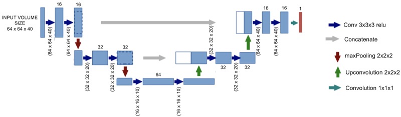
All dynamic 18F-FET PET brain image volumes were temporally realigned to the first dynamic acquisition, coregistered and spatially normalized onto the Montreal Neurological Institute template. Ground truth segmentations were obtained using manual delineation and thresholding (1.3 x background). The volumetric CNN was implemented based on a modified Keras implementation of a U-Net library with 3 layers for the encoding and decoding paths. Dice similarity coefficient (DSC) was used as an accuracy measure of segmentation.

RESULTS

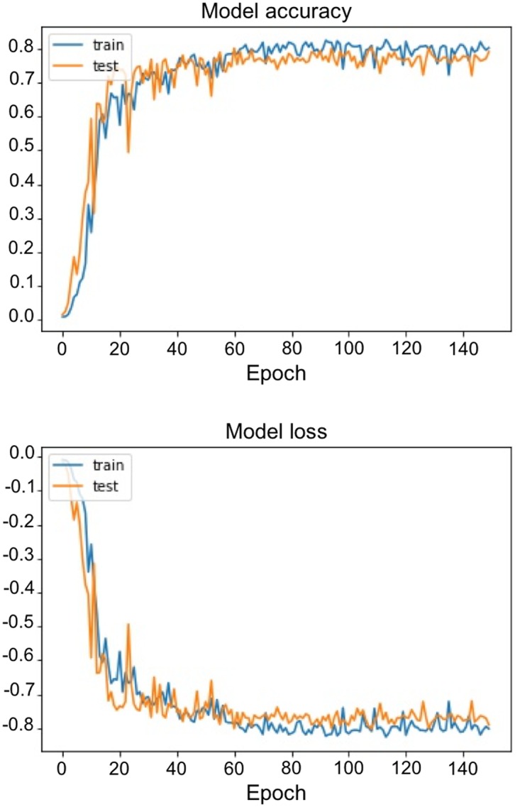
Thirty-seven patients were included (26 [70%] in the training set and 11 [30%] in the validation set). All 11 lesions were accurately detected with no false positive, resulting in a sensitivity and a specificity for the detection at the tumor level of 100%. After 150 epochs, DSC reached 0.7924 in the training set and 0.7911 in the validation set. After morphological dilatation and fixed thresholding of the predicted U-Net mask a substantial improvement of the DSC to 0.8231 (+ 4.1%) was noted. At the voxel level, this segmentation led to a 0.88 sensitivity [95% CI, 87.1 to, 88.2%] a 0.99 specificity [99.9 to 99.9%], a 0.78 positive predictive value: [76.9 to 78.3%], and a 0.99 negative predictive value [99.9 to 99.9%].

CONCLUSIONS

With relatively high performance, it was proposed the first full 3D automated procedure for segmentation of 18F-FET PET brain images of patients with different gliomas using a U-Net CNN architecture.

摘要

简介

氨基酸正电子发射断层扫描(PET)越来越多地用于胶质瘤患者的诊断,包括鉴别诊断、肿瘤扩展评估、治疗计划和随访。最近,计算机视觉和机器学习的进展已经应用于医学成像。目的是证明基于全 3D U-Net 卷积神经网络(CNN)的自动 18F-氟乙基酪氨酸(18F-FET)PET 病变检测和分割的可行性。

方法

所有动态 18F-FET PET 脑图像体积均进行时间校正,以获得第一个动态采集的图像,与蒙特利尔神经学研究所模板进行配准,并进行空间标准化。使用手动勾画和阈值(1.3x 背景)获得地面真实分割。体积 CNN 是基于带有 3 个编码和解码路径层的 U-Net 库的修改后的 Keras 实现来实现的。Dice 相似系数(DSC)用作分割准确性的度量。

结果

共纳入 37 例患者(训练集 26 例[70%],验证集 11 例[30%])。所有 11 个病变均准确检测到,无假阳性,肿瘤水平的灵敏度和特异性均为 100%。在 150 个时期后,训练集的 DSC 达到 0.7924,验证集的 DSC 达到 0.7911。在预测 U-Net 掩模的形态扩张和固定阈值之后,DSC 显著提高到 0.8231(+4.1%)。在体素水平上,该分割方法的灵敏度为 0.88[95%CI,87.1%至 88.2%],特异性为 0.99[99.9%至 99.9%],阳性预测值为 0.78[76.9%至 78.3%],阴性预测值为 0.99[99.9%至 99.9%]。

结论

使用 U-Net CNN 架构,提出了首个用于不同脑胶质瘤患者 18F-FET PET 脑图像分割的全自动全 3D 处理程序,具有较高的性能。

https://cdn.ncbi.nlm.nih.gov/pmc/blobs/72cf/5898737/25de5af58369/pone.0195798.g003.jpg
https://cdn.ncbi.nlm.nih.gov/pmc/blobs/72cf/5898737/496d08799b08/pone.0195798.g001.jpg
https://cdn.ncbi.nlm.nih.gov/pmc/blobs/72cf/5898737/370ff80f2815/pone.0195798.g002.jpg
https://cdn.ncbi.nlm.nih.gov/pmc/blobs/72cf/5898737/25de5af58369/pone.0195798.g003.jpg
https://cdn.ncbi.nlm.nih.gov/pmc/blobs/72cf/5898737/496d08799b08/pone.0195798.g001.jpg
https://cdn.ncbi.nlm.nih.gov/pmc/blobs/72cf/5898737/370ff80f2815/pone.0195798.g002.jpg
https://cdn.ncbi.nlm.nih.gov/pmc/blobs/72cf/5898737/25de5af58369/pone.0195798.g003.jpg

相似文献

1
Automatic lesion detection and segmentation of 18F-FET PET in gliomas: A full 3D U-Net convolutional neural network study.基于全 3D U-Net 卷积神经网络的脑胶质瘤 18F-FET PET 自动病灶检测与分割研究。
PLoS One. 2018 Apr 13;13(4):e0195798. doi: 10.1371/journal.pone.0195798. eCollection 2018.
2
A multi-label CNN model for the automatic detection and segmentation of gliomas using [F]FET PET imaging.一种基于多标签卷积神经网络的自动检测和分割方法,用于使用 [F]FET PET 成像检测脑胶质瘤。
Eur J Nucl Med Mol Imaging. 2023 Jul;50(8):2441-2452. doi: 10.1007/s00259-023-06193-5. Epub 2023 Mar 18.
3
Voxel-based 18F-FET PET segmentation and automatic clustering of tumor voxels: A significant association with IDH1 mutation status and survival in patients with gliomas.基于体素的 18F-FET PET 分割和肿瘤体素的自动聚类:与 IDH1 突变状态和胶质瘤患者生存的显著关联。
PLoS One. 2018 Jun 28;13(6):e0199379. doi: 10.1371/journal.pone.0199379. eCollection 2018.
4
Assessment of various strategies for 18F-FET PET-guided delineation of target volumes in high-grade glioma patients.评估18F-FET PET引导下高级别胶质瘤患者靶区勾画的各种策略。
Eur J Nucl Med Mol Imaging. 2009 Feb;36(2):182-93. doi: 10.1007/s00259-008-0943-6. Epub 2008 Sep 26.
5
Two-stage deep learning model for fully automated pancreas segmentation on computed tomography: Comparison with intra-reader and inter-reader reliability at full and reduced radiation dose on an external dataset.基于 CT 的全自动胰腺分割的两阶段深度学习模型:在外部数据集上比较全剂量和低剂量下的同读者和异读者可靠性。
Med Phys. 2021 May;48(5):2468-2481. doi: 10.1002/mp.14782. Epub 2021 Mar 16.
6
Automated glioma grading on conventional MRI images using deep convolutional neural networks.使用深度卷积神经网络对传统MRI图像进行自动脑胶质瘤分级
Med Phys. 2020 Jul;47(7):3044-3053. doi: 10.1002/mp.14168. Epub 2020 May 11.
7
Serial 18F-FET PET Imaging of Primarily 18F-FET-Negative Glioma: Does It Make Sense?原发性18F-FET阴性胶质瘤的连续18F-FET PET成像:有意义吗?
J Nucl Med. 2016 Aug;57(8):1177-82. doi: 10.2967/jnumed.115.171033. Epub 2016 Mar 31.
8
An interindividual comparison of O-(2-[18F]fluoroethyl)-L-tyrosine (FET)- and L-[methyl-11C]methionine (MET)-PET in patients with brain gliomas and metastases.个体间比较 O-(2-[18F]氟乙基)-L-酪氨酸(FET)和 L-[甲基-11C]蛋氨酸(MET)-PET 在脑胶质瘤和转移瘤患者中的应用。
Int J Radiat Oncol Biol Phys. 2011 Nov 15;81(4):1049-58. doi: 10.1016/j.ijrobp.2010.07.002. Epub 2011 May 11.
9
Postoperative glioma segmentation in CT image using deep feature fusion model guided by multi-sequence MRIs.基于多序列 MRI 引导的深度特征融合模型的 CT 图像术后脑肿瘤分割。
Eur Radiol. 2020 Feb;30(2):823-832. doi: 10.1007/s00330-019-06441-z. Epub 2019 Oct 24.
10
An investigation of the effect of fat suppression and dimensionality on the accuracy of breast MRI segmentation using U-nets.利用 U-Nets 研究脂肪抑制和维度对乳腺 MRI 分割准确性的影响。
Med Phys. 2019 Mar;46(3):1230-1244. doi: 10.1002/mp.13375. Epub 2019 Feb 4.

引用本文的文献

1
Artificial intelligence in medicine: Current applications in cardiology, oncology, and radiology.医学中的人工智能:当前在心脏病学、肿瘤学和放射学中的应用。
World J Methodol. 2025 Dec 20;15(4):106854. doi: 10.5662/wjm.v15.i4.106854.
2
F-FDG PET-based liver segmentation using deep-learning.基于深度学习的F-FDG正电子发射断层扫描肝脏分割
Phys Eng Sci Med. 2025 Jul 15. doi: 10.1007/s13246-025-01595-1.
3
Systematic double reading for oncological PET/CT scans: insights from a prospective multicentre study in 678 patients.肿瘤PET/CT扫描的系统双人阅片:来自一项针对678例患者的前瞻性多中心研究的见解

本文引用的文献

1
O-(2-[F]fluoroethyl)-L-tyrosine PET in gliomas: influence of data processing in different centres.O-(2-[F]氟乙基)-L-酪氨酸PET在胶质瘤中的应用:不同中心数据处理的影响
EJNMMI Res. 2017 Aug 16;7(1):64. doi: 10.1186/s13550-017-0316-x.
2
Deep Learning in Medical Image Analysis.医学图像分析中的深度学习
Annu Rev Biomed Eng. 2017 Jun 21;19:221-248. doi: 10.1146/annurev-bioeng-071516-044442. Epub 2017 Mar 9.
3
Classification and evaluation strategies of auto-segmentation approaches for PET: Report of AAPM task group No. 211.
EJNMMI Rep. 2025 Jun 13;9(1):19. doi: 10.1186/s41824-025-00253-9.
4
Interobserver ground-truth variability limits performance of automated glioblastoma segmentation on [F]FET PET.观察者间的真实情况变异性限制了[F]FET PET上胶质母细胞瘤自动分割的性能。
EJNMMI Phys. 2025 Jun 6;12(1):54. doi: 10.1186/s40658-025-00767-y.
5
Characterization of artificial intelligence performance for lesion detection using synthetic lesions in PET imaging.利用PET成像中的合成病变对人工智能病变检测性能进行表征。
Med Phys. 2025 Jun;52(6):3994-4007. doi: 10.1002/mp.17694. Epub 2025 Feb 24.
6
A privacy-preserved horizontal federated learning for malignant glioma tumour detection using distributed data-silos.一种使用分布式数据孤岛进行恶性胶质瘤肿瘤检测的隐私保护水平联邦学习。
PLoS One. 2025 Feb 11;20(2):e0316543. doi: 10.1371/journal.pone.0316543. eCollection 2025.
7
Brain CT image classification based on mask RCNN and attention mechanism.基于Mask R-CNN和注意力机制的脑CT图像分类
Sci Rep. 2024 Nov 26;14(1):29300. doi: 10.1038/s41598-024-78566-1.
8
Automatic detection and delineation of pediatric gliomas on combined [F]FET PET and MRI.基于联合[F]FET正电子发射断层显像(PET)与磁共振成像(MRI)的儿童胶质瘤自动检测与勾画
Front Nucl Med. 2022 Aug 24;2:960820. doi: 10.3389/fnume.2022.960820. eCollection 2022.
9
Semi-supervised learning towards automated segmentation of PET images with limited annotations: application to lymphoma patients.半监督学习在有限标注 PET 图像自动分割中的应用:在淋巴瘤患者中的应用。
Phys Eng Sci Med. 2024 Sep;47(3):833-849. doi: 10.1007/s13246-024-01408-x. Epub 2024 Mar 21.
10
Need for Objective Task-Based Evaluation of Image Segmentation Algorithms for Quantitative PET: A Study with ACRIN 6668/RTOG 0235 Multicenter Clinical Trial Data.客观基于任务的定量 PET 图像分割算法评估的必要性:一项使用 ACRIN 6668/RTOG 0235 多中心临床试验数据的研究。
J Nucl Med. 2024 Mar 1;65(3):485-92. doi: 10.2967/jnumed.123.266018. Epub 2024 Feb 15.
自动分割方法在正电子发射断层扫描中的分类和评估策略:AAPM 工作组第 211 号报告。
Med Phys. 2017 Jun;44(6):e1-e42. doi: 10.1002/mp.12124. Epub 2017 May 18.
4
Dynamic O-(2-18F-fluoroethyl)-L-tyrosine positron emission tomography differentiates brain metastasis recurrence from radiation injury after radiotherapy.动态O-(2-¹⁸F-氟乙基)-L-酪氨酸正电子发射断层扫描可区分脑转移瘤复发与放疗后的放射性损伤。
Neuro Oncol. 2017 Feb 1;19(2):281-288. doi: 10.1093/neuonc/now149.
5
Prognostic Value of O-(2-[18F]-Fluoroethyl)-L-Tyrosine-Positron Emission Tomography Imaging for Histopathologic Characteristics and Progression-Free Survival in Patients with Low-Grade Glioma.O-(2-[18F]-氟乙基)-L-酪氨酸-正电子发射断层扫描成像对低级别胶质瘤患者组织病理学特征及无进展生存期的预后价值
World Neurosurg. 2016 May;89:230-9. doi: 10.1016/j.wneu.2016.01.085. Epub 2016 Mar 9.
6
Volumetric Analysis of F-18-FET-PET Imaging for Brain Metastases.用于脑转移瘤的F-18-FET-PET成像的容积分析
World Neurosurg. 2015 Dec;84(6):1790-7. doi: 10.1016/j.wneu.2015.07.067. Epub 2015 Aug 6.
7
Performance of 18F-FET versus 18F-FDG-PET for the diagnosis and grading of brain tumors: systematic review and meta-analysis.18F-FET与18F-FDG-PET在脑肿瘤诊断和分级中的性能:系统评价与荟萃分析
Neuro Oncol. 2016 Mar;18(3):426-34. doi: 10.1093/neuonc/nov148. Epub 2015 Aug 4.
8
Pretherapy metabolic tumour volume is an independent predictor of outcome in patients with diffuse large B-cell lymphoma.治疗前代谢肿瘤体积是弥漫性大B细胞淋巴瘤患者预后的独立预测指标。
Eur J Nucl Med Mol Imaging. 2014 Nov;41(11):2017-22. doi: 10.1007/s00259-014-2822-7. Epub 2014 Jun 6.
9
Machine learning for neuroimaging with scikit-learn.使用 scikit-learn 进行神经影像学的机器学习。
Front Neuroinform. 2014 Feb 21;8:14. doi: 10.3389/fninf.2014.00014. eCollection 2014.
10
Molecular neuro-oncology in clinical practice: a new horizon.分子神经肿瘤学在临床实践中的应用:新的前景。
Lancet Oncol. 2013 Aug;14(9):e370-9. doi: 10.1016/S1470-2045(13)70168-2.