Raspini Gregorio, Wolff Jan, Helminen Mika, Raspini Giovacchino, Raspini Mario, Sándor George K
Department of Oral and Maxillofacial Surgery, University of Oulu, OuluFinland.
Department of Oral and Maxillofacial Surgery/Pathology & 3D Innovation Lab, VU University Medical Center, AmsterdamThe Netherlands.
J Oral Maxillofac Res. 2018 Mar 31;9(1):e2. doi: 10.5037/jomr.2018.9102. eCollection 2018 Jan-Mar.
The aim of this study was to assess the interaction of a bioactive glass scaffold with cells derived from dental pulp, dental follicle and periodontal ligament.
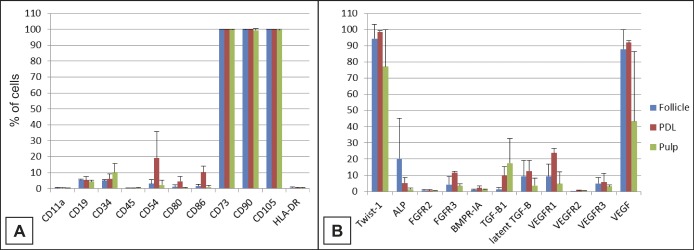
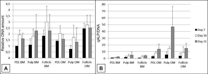
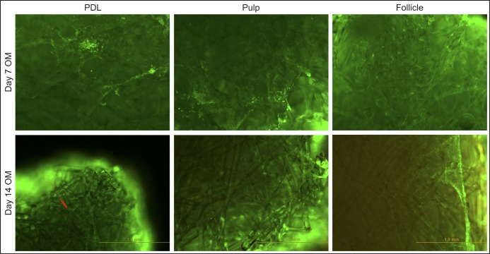
Impacted third molars were surgically removed from three young donors. Cells from the dental pulp, follicle and periodontal ligament tissues were isolated and expanded. Different cell populations were characterised using specific CD markers. Expanded pulp, follicle and periodontal cells were then seeded onto bioactive glass scaffolds and cultured in osteogenic medium or basic medium. Cell attachment, viability, proliferation and alkaline phosphatase activity were assessed.
This study revealed good biocompatibility of the specific bioactive glass configuration tested and the osteogenic induction of cells derived from dental pulp, dental follicle and periodontal ligament. Osteogenic medium seemed to increase the differentiation pattern and dental pulp stem cells showed the most positive results compared to periodontal ligament and dental follicle stem cells.
Dental pulp stem cells combined with a bioactive glass scaffold and exposed to osteogenic medium represent a promising combination for future study of hard tissue regeneration in the cranio-maxillofacial skeleton.
本研究旨在评估生物活性玻璃支架与牙髓、牙囊和牙周膜来源细胞之间的相互作用。
从三名年轻供体手术拔除阻生第三磨牙。分离并扩增牙髓、牙囊和牙周膜组织中的细胞。使用特异性CD标志物对不同细胞群体进行表征。然后将扩增后的牙髓、牙囊和牙周细胞接种到生物活性玻璃支架上,并在成骨培养基或基础培养基中培养。评估细胞附着、活力、增殖和碱性磷酸酶活性。
本研究揭示了所测试的特定生物活性玻璃构型具有良好的生物相容性,以及对牙髓、牙囊和牙周膜来源细胞的成骨诱导作用。成骨培养基似乎增加了分化模式,与牙周膜和牙囊干细胞相比,牙髓干细胞显示出最积极的结果。
牙髓干细胞与生物活性玻璃支架结合并暴露于成骨培养基中,是未来颅颌面骨骼硬组织再生研究的一个有前景的组合。