Max Planck Institute for Human Development Center for Lifespan Psychology Berlin Germany.
Center for Military Mental Health Military Hospital Berlin Berlin Germany.
Brain Behav. 2018 Apr 6;8(5):e00956. doi: 10.1002/brb3.956. eCollection 2018 May.
Smaller hippocampal volumes are one of the most consistent findings in neuroimaging studies of post-traumatic stress disorder (PTSD). However, very few prospective studies have assessed changes in hippocampal gray matter prior to and following therapy for PTSD, and no neuroimaging studies to date have longitudinally assessed military populations.
A pilot study was conducted, assessing patients with combat-related PTSD with structural MRI. Participants were then assigned either to a treatment group or waiting-list control group. After the treatment group received multimodal psychological therapy for approximately 6 weeks, both groups completed a second neuroimaging assessment.
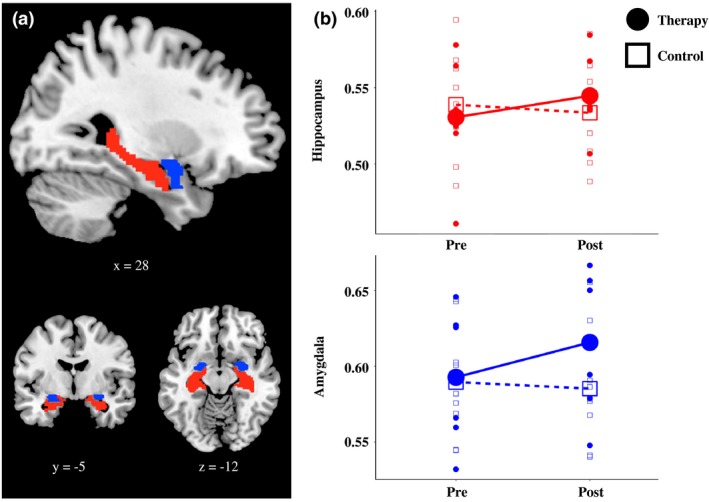
Region-of-interest analysis was used to measure gray matter volume in the hippocampus and amygdala. There was a group by time interaction; the therapy group ( = 6) showed a significant increase in hippocampal volume and a nonsignificant trend toward an increase in amygdala volume following therapy, while no change was observed in the waiting-list group ( = 9).
This study provides initial evidence for increases in gray matter volume in the hippocampus in response to therapy for combat-related PTSD.
创伤后应激障碍(PTSD)的神经影像学研究发现,较小的海马体体积是最一致的发现之一。然而,很少有前瞻性研究在 PTSD 治疗前后评估海马灰质的变化,迄今为止也没有神经影像学研究对军事人群进行纵向评估。
进行了一项试点研究,对与战斗相关的 PTSD 患者进行结构 MRI 评估。然后,参与者被分配到治疗组或候补名单对照组。在治疗组接受了大约 6 周的多模式心理治疗后,两组都完成了第二次神经影像学评估。
采用感兴趣区域分析测量海马体和杏仁核的灰质体积。存在组间时间交互作用;治疗组(n=6)在治疗后海马体体积显著增加,杏仁核体积有增加的趋势但无统计学意义,而候补名单组(n=9)无变化。
这项研究初步提供了证据,表明针对与战斗相关的 PTSD 的治疗可引起海马体灰质体积增加。