Khalvati Farzad, Zhang Junjie, Chung Audrey G, Shafiee Mohammad Javad, Wong Alexander, Haider Masoom A
Department of Medical Imaging, University of Toronto and Sunnybrook Research Institute, Toronto, Ontario, Canada.
Department of Systems Design Engineering, University of Waterloo, Waterloo, Ontario, Canada.
BMC Med Imaging. 2018 May 16;18(1):16. doi: 10.1186/s12880-018-0258-4.
Quantitative radiomic features provide a plethora of minable data extracted from multi-parametric magnetic resonance imaging (MP-MRI) which can be used for accurate detection and localization of prostate cancer. While most cancer detection algorithms utilize either voxel-based or region-based feature models, the complexity of prostate tumour phenotype in MP-MRI requires a more sophisticated framework to better leverage available data and exploit a priori knowledge in the field.
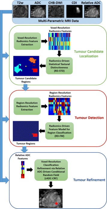
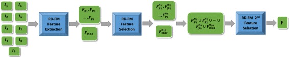
In this paper, we present MPCaD, a novel Multi-scale radiomics-driven framework for Prostate Cancer Detection and localization which leverages radiomic feature models at different scales as well as incorporates a priori knowledge of the field. Tumour candidate localization is first performed using a statistical texture distinctiveness strategy that leverages a voxel-resolution feature model to localize tumour candidate regions. Tumour region classification via a region-resolution feature model is then performed to identify tumour regions. Both voxel-resolution and region-resolution feature models are built upon and extracted from six different MP-MRI modalities. Finally, a conditional random field framework that is driven by voxel-resolution relative ADC features is used to further refine the localization of the tumour regions in the peripheral zone to improve the accuracy of the results.
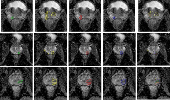
The proposed framework is evaluated using clinical prostate MP-MRI data from 30 patients, and results demonstrate that the proposed framework exhibits enhanced separability of cancerous and healthy tissue, as well as outperforms individual quantitative radiomics models for prostate cancer detection.
Quantitative radiomic features extracted from MP-MRI of prostate can be utilized to detect and localize prostate cancer.
定量放射组学特征可提供从多参数磁共振成像(MP-MRI)中提取的大量可挖掘数据,这些数据可用于前列腺癌的准确检测和定位。虽然大多数癌症检测算法使用基于体素或基于区域的特征模型,但MP-MRI中前列腺肿瘤表型的复杂性需要一个更复杂的框架,以更好地利用现有数据并利用该领域的先验知识。
在本文中,我们提出了MPCaD,这是一种用于前列腺癌检测和定位的新型多尺度放射组学驱动框架,它利用不同尺度的放射组学特征模型,并结合了该领域的先验知识。首先使用统计纹理独特性策略进行肿瘤候选定位,该策略利用体素分辨率特征模型定位肿瘤候选区域。然后通过区域分辨率特征模型进行肿瘤区域分类,以识别肿瘤区域。体素分辨率和区域分辨率特征模型均基于六种不同的MP-MRI模态构建并从中提取。最后,使用由体素分辨率相对ADC特征驱动的条件随机场框架进一步细化外周区肿瘤区域的定位,以提高结果的准确性。
使用来自30名患者的临床前列腺MP-MRI数据对所提出的框架进行评估,结果表明所提出的框架在癌组织和健康组织之间表现出更强的可分离性,并且在前列腺癌检测方面优于单个定量放射组学模型。
从前列腺MP-MRI中提取的定量放射组学特征可用于检测和定位前列腺癌。