• 文献检索
  • 文档翻译
  • 深度研究
  • 学术资讯
  • Suppr Zotero 插件Zotero 插件
  • 邀请有礼
  • 套餐&价格
  • 历史记录
应用&插件
Suppr Zotero 插件Zotero 插件浏览器插件Mac 客户端Windows 客户端微信小程序
定价
高级版会员购买积分包购买API积分包
服务
文献检索文档翻译深度研究API 文档MCP 服务
关于我们
关于 Suppr公司介绍联系我们用户协议隐私条款
关注我们

Suppr 超能文献

核心技术专利:CN118964589B侵权必究
粤ICP备2023148730 号-1Suppr @ 2026

文献检索

告别复杂PubMed语法,用中文像聊天一样搜索,搜遍4000万医学文献。AI智能推荐,让科研检索更轻松。

立即免费搜索

文件翻译

保留排版,准确专业,支持PDF/Word/PPT等文件格式,支持 12+语言互译。

免费翻译文档

深度研究

AI帮你快速写综述,25分钟生成高质量综述,智能提取关键信息,辅助科研写作。

立即免费体验

测定活细胞中 CD59 的膜环境。

Determination of the Membrane Environment of CD59 in Living Cells.

机构信息

Institute of Applied Physics, TU Wien, Wiedner Hauptstrasse 8-10, Vienna 1040, Austria.

出版信息

Biomolecules. 2018 May 17;8(2):28. doi: 10.3390/biom8020028.

DOI:10.3390/biom8020028
PMID:29772810
原文链接:https://pmc.ncbi.nlm.nih.gov/articles/PMC6023084/
Abstract

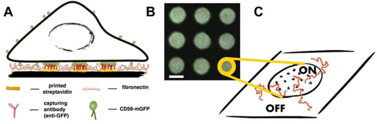
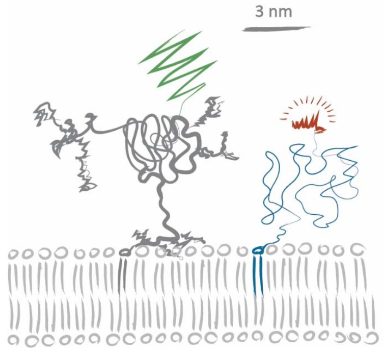
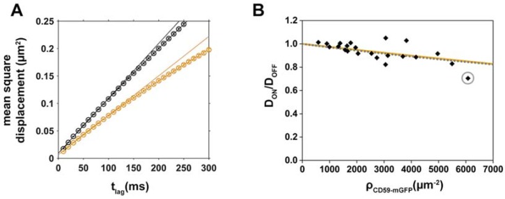
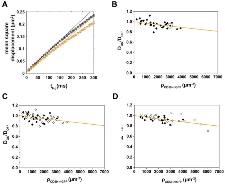
The organization and dynamics of proteins and lipids in the plasma membrane, and their role in membrane functionality, have been subject of a long-lasting debate. Specifically, it is unclear to what extent membrane proteins are affected by their immediate lipid environment and vice versa. Studies on model membranes and plasma membrane vesicles indicated preferences of proteins for lipid phases characterized by different acyl chain order; however, whether such phases do indeed exist in live cells is still not known. Here, we refine a previously developed micropatterning approach combined with single molecule tracking to quantify the influence of the glycosylphosphatidylinositol-anchored (GPI-anchored) protein CD59 on its molecular environment directly in the live cell plasma membrane. We find that locally enriched and immobilized CD59 presents obstacles to the diffusion of fluorescently labeled lipids with a different phase-partitioning behavior independent of cell cholesterol levels and type of lipid. Our results give no evidence for either specific binding of the lipids to CD59 or the existence of nanoscopic ordered membrane regions associated with CD59.

摘要

质膜中蛋白质和脂质的组织和动力学及其在膜功能中的作用一直是一个长期存在的争论。具体来说,膜蛋白在多大程度上受到其直接脂质环境的影响,反之亦然,这一点还不清楚。关于模型膜和质膜小泡的研究表明,蛋白质对不同酰链有序性特征的脂质相具有偏好性;然而,在活细胞中是否确实存在这样的相仍然未知。在这里,我们改进了以前开发的一种微图案化方法,结合单分子跟踪,以直接在活细胞质膜中定量测定糖基磷脂酰肌醇锚定(GPI 锚定)蛋白 CD59 对其分子环境的影响。我们发现,局部富集和固定的 CD59 会阻碍具有不同相分离行为的荧光标记脂质的扩散,而与细胞胆固醇水平和脂质类型无关。我们的结果没有证据表明脂质与 CD59 有特异性结合,也没有证据表明与 CD59 相关的纳米级有序膜区域的存在。

https://cdn.ncbi.nlm.nih.gov/pmc/blobs/4d1f/6023084/5f06f3618f71/biomolecules-08-00028-g004.jpg
https://cdn.ncbi.nlm.nih.gov/pmc/blobs/4d1f/6023084/dbe3ccdd2f5a/biomolecules-08-00028-g001.jpg
https://cdn.ncbi.nlm.nih.gov/pmc/blobs/4d1f/6023084/c96ba9b9ead7/biomolecules-08-00028-g002.jpg
https://cdn.ncbi.nlm.nih.gov/pmc/blobs/4d1f/6023084/5c6b426a6fae/biomolecules-08-00028-g003.jpg
https://cdn.ncbi.nlm.nih.gov/pmc/blobs/4d1f/6023084/5f06f3618f71/biomolecules-08-00028-g004.jpg

相似文献

1
Determination of the Membrane Environment of CD59 in Living Cells.测定活细胞中 CD59 的膜环境。
Biomolecules. 2018 May 17;8(2):28. doi: 10.3390/biom8020028.
2
GPI-anchored proteins do not reside in ordered domains in the live cell plasma membrane.糖基磷脂酰肌醇(GPI)锚定蛋白并不存在于活细胞质膜的有序结构域中。
Nat Commun. 2015 Apr 21;6:6969. doi: 10.1038/ncomms7969.
3
Revealing the Raft Domain Organization in the Plasma Membrane by Single-Molecule Imaging of Fluorescent Ganglioside Analogs.通过荧光神经节苷脂类似物的单分子成像揭示质膜中的筏域组织
Methods Enzymol. 2018;598:267-282. doi: 10.1016/bs.mie.2017.06.038. Epub 2017 Oct 31.
4
Dynamics of raft molecules in the cell and artificial membranes: approaches by pulse EPR spin labeling and single molecule optical microscopy.细胞及人工膜中筏分子的动力学:脉冲电子顺磁共振自旋标记和单分子光学显微镜方法
Biochim Biophys Acta. 2003 Mar 10;1610(2):231-43. doi: 10.1016/s0005-2736(03)00021-x.
5
Single-molecule microscopy reveals heterogeneous dynamics of lipid raft components upon TCR engagement.单分子显微镜技术揭示了T细胞受体(TCR)激活后脂筏成分的异质性动力学。
Int Immunol. 2007 May;19(5):675-84. doi: 10.1093/intimm/dxm032. Epub 2007 Apr 19.
6
Unraveling of Lipid Raft Organization in Cell Plasma Membranes by Single-Molecule Imaging of Ganglioside Probes.通过神经节苷脂探针的单分子成像揭示细胞质膜中脂筏组织的解聚。
Adv Exp Med Biol. 2018;1104:41-58. doi: 10.1007/978-981-13-2158-0_3.
7
Partitioning, diffusion, and ligand binding of raft lipid analogs in model and cellular plasma membranes.筏脂类似物在模型细胞膜和细胞质膜中的分区、扩散及配体结合
Biochim Biophys Acta. 2012 Jul;1818(7):1777-84. doi: 10.1016/j.bbamem.2012.03.007.
8
Development of new ganglioside probes and unraveling of raft domain structure by single-molecule imaging.新型神经节苷脂探针的开发及利用单分子成像技术揭示脂筏结构
Biochim Biophys Acta Gen Subj. 2017 Oct;1861(10):2494-2506. doi: 10.1016/j.bbagen.2017.07.012. Epub 2017 Jul 20.
9
Determination of lipid raft partitioning of fluorescently-tagged probes in living cells by Fluorescence Correlation Spectroscopy (FCS).通过荧光相关光谱法(FCS)测定活细胞中荧光标记探针的脂筏分配情况。
J Vis Exp. 2012 Apr 6(62):e3513. doi: 10.3791/3513.
10
(Un)confined diffusion of CD59 in the plasma membrane determined by high-resolution single molecule microscopy.通过高分辨率单分子显微镜测定CD59在质膜中的(非)受限扩散。
Biophys J. 2007 May 15;92(10):3719-28. doi: 10.1529/biophysj.106.095398. Epub 2007 Feb 26.

引用本文的文献

1
Soft lithography-based biomolecule patterning techniques and their applications in subcellular protein interaction analysis.基于软光刻的生物分子图案化技术及其在亚细胞蛋白质相互作用分析中的应用。
Mater Today Bio. 2025 Mar 17;32:101672. doi: 10.1016/j.mtbio.2025.101672. eCollection 2025 Jun.
2
Measurement of FGFR3 signaling at the cell membrane via total internal reflection fluorescence microscopy to compare the activation of FGFR3 mutants.通过全内反射荧光显微镜测量细胞膜上 FGFR3 信号,以比较 FGFR3 突变体的激活情况。
J Biol Chem. 2023 Feb;299(2):102832. doi: 10.1016/j.jbc.2022.102832. Epub 2022 Dec 27.
3
A micropatterning platform for quantifying interaction kinetics between the T cell receptor and an intracellular binding protein.

本文引用的文献

1
Monte Carlo simulations of protein micropatterning in biomembranes: effects of immobile sticky obstacles.生物膜中蛋白质微图案化的蒙特卡罗模拟:固定粘性障碍物的影响。
J Phys D Appl Phys. 2016 Aug 9;49(36). doi: 10.1088/0022-3727/49/36/364002.
2
Nanoscale Membrane Budding Induced by CTxB and Detected via Polarized Localization Microscopy.由霍乱毒素B亚基(CTxB)诱导并通过偏振定位显微镜检测的纳米级膜出芽
Biophys J. 2017 Oct 17;113(8):1795-1806. doi: 10.1016/j.bpj.2017.08.031.
3
Diffusion of lipids and GPI-anchored proteins in actin-free plasma membrane vesicles measured by STED-FCS.
一种用于量化 T 细胞受体与细胞内结合蛋白之间相互作用动力学的微图案化平台。
Sci Rep. 2019 Mar 1;9(1):3288. doi: 10.1038/s41598-019-39865-0.
4
A Fast and Simple Contact Printing Approach to Generate 2D Protein Nanopatterns.一种用于生成二维蛋白质纳米图案的快速简便的接触印刷方法。
Front Chem. 2019 Jan 24;6:655. doi: 10.3389/fchem.2018.00655. eCollection 2018.
5
Tunable DNA Hybridization Enables Spatially and Temporally Controlled Surface-Anchoring of Biomolecular Cargo.可调谐 DNA 杂交实现了生物分子货物的空间和时间控制的表面锚定。
Langmuir. 2018 Dec 11;34(49):15021-15027. doi: 10.1021/acs.langmuir.8b01942. Epub 2018 Aug 30.
通过受激发射损耗荧光相关光谱法(STED-FCS)测量无肌动蛋白质膜囊泡中脂质和糖基磷脂酰肌醇锚定蛋白的扩散。
Mol Biol Cell. 2017 Jun 1;28(11):1507-1518. doi: 10.1091/mbc.E16-07-0536. Epub 2017 Apr 12.
4
Protein Micropatterning Assay: Quantitative Analysis of Protein-Protein Interactions.蛋白质微图案分析:蛋白质 - 蛋白质相互作用的定量分析
Methods Mol Biol. 2017;1550:261-270. doi: 10.1007/978-1-4939-6747-6_18.
5
Raft-based interactions of gangliosides with a GPI-anchored receptor.神经节苷脂与糖基磷脂酰肌醇锚定受体的筏基相互作用。
Nat Chem Biol. 2016 Jun;12(6):402-10. doi: 10.1038/nchembio.2059. Epub 2016 Apr 4.
6
A comparative study on fluorescent cholesterol analogs as versatile cellular reporters.荧光胆固醇类似物作为多功能细胞报告分子的比较研究
J Lipid Res. 2016 Feb;57(2):299-309. doi: 10.1194/jlr.M065326. Epub 2015 Dec 23.
7
With or without rafts? Alternative views on cell membranes.有脂筏还是没有脂筏?关于细胞膜的不同观点。
Bioessays. 2016 Feb;38(2):129-39. doi: 10.1002/bies.201500150. Epub 2015 Dec 15.
8
Transbilayer lipid interactions mediate nanoclustering of lipid-anchored proteins.跨膜脂质相互作用介导脂质锚定蛋白的纳米簇集。
Cell. 2015 Apr 23;161(3):581-594. doi: 10.1016/j.cell.2015.03.048.
9
GPI-anchored proteins do not reside in ordered domains in the live cell plasma membrane.糖基磷脂酰肌醇(GPI)锚定蛋白并不存在于活细胞质膜的有序结构域中。
Nat Commun. 2015 Apr 21;6:6969. doi: 10.1038/ncomms7969.
10
Scanning STED-FCS reveals spatiotemporal heterogeneity of lipid interaction in the plasma membrane of living cells.扫描 STED-FCS 揭示了活细胞质膜中脂质相互作用的时空异质性。
Nat Commun. 2014 Nov 20;5:5412. doi: 10.1038/ncomms6412.