Toronto General Hospital Research Institute, Division of Cardiovascular Surgery, University Health Network, Toronto, Ontario, Canada.
Department of Surgery, Division of Cardiac Surgery, University of Toronto, Toronto, Ontario, Canada.
Theranostics. 2018 Apr 9;8(10):2752-2764. doi: 10.7150/thno.22599. eCollection 2018.
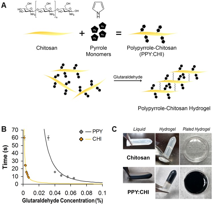
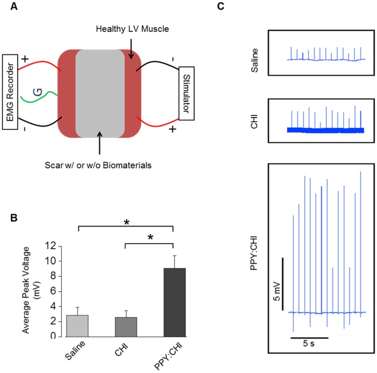
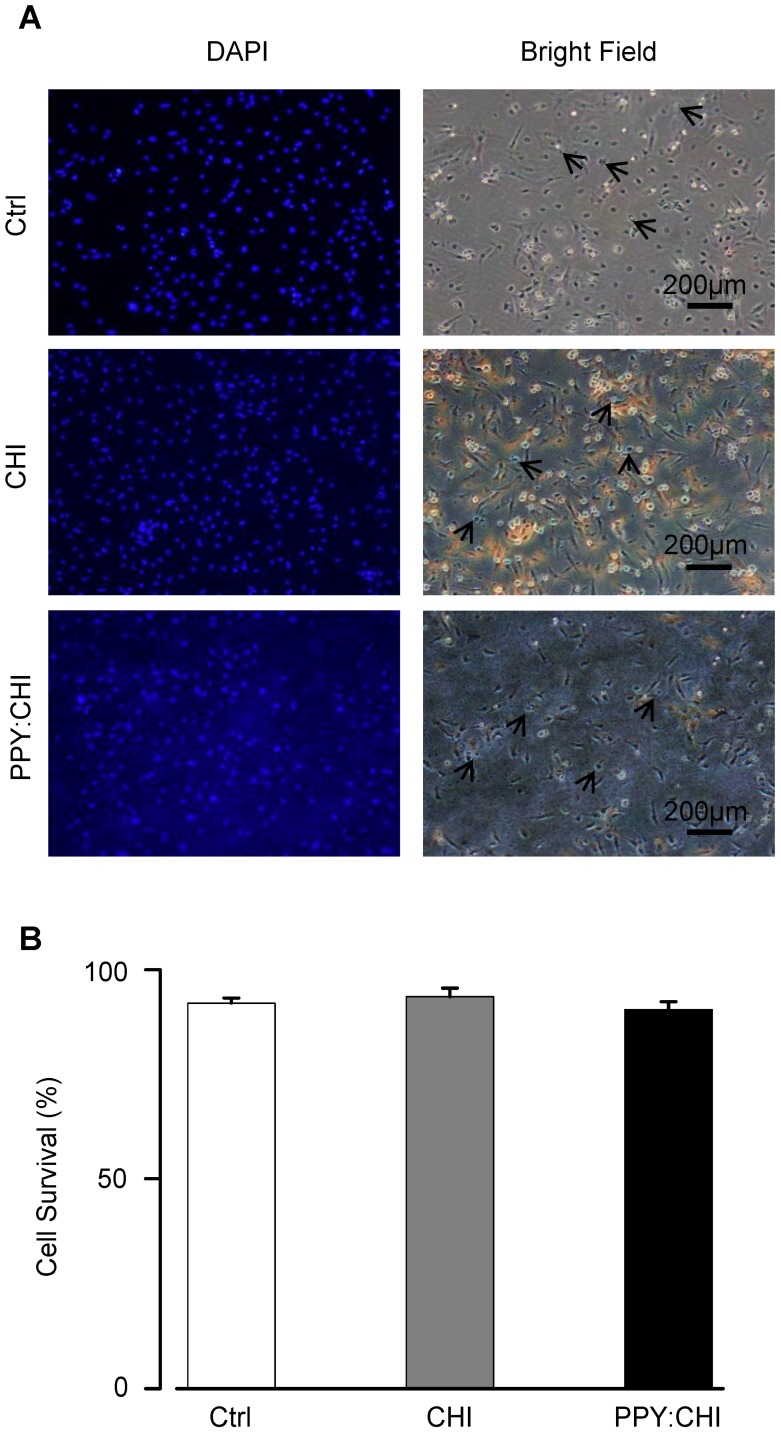
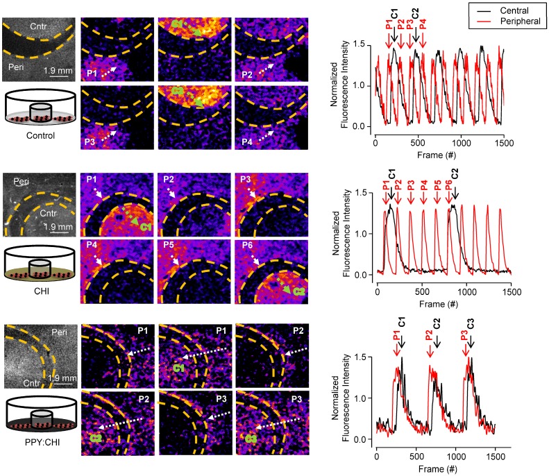
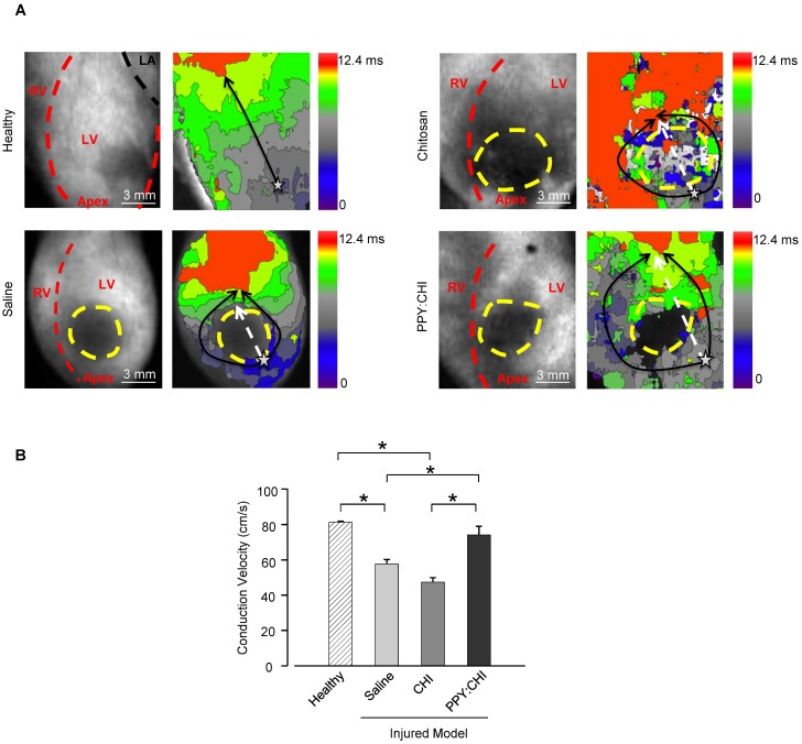
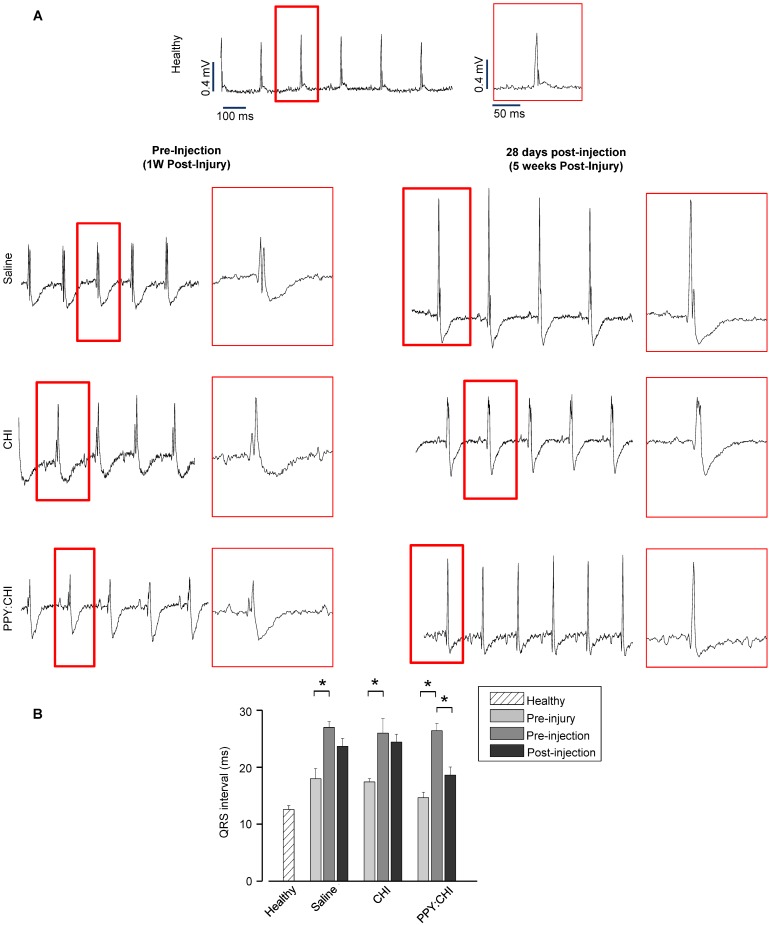
The post-myocardial infarction (MI) scar interrupts electrical impulse propagation and delays regional contraction, which contributes to ventricular dysfunction. We investigated the potential of an injectable conductive biomaterial to restore scar tissue conductivity and re-establish synchronous ventricular contraction. A conductive biomaterial was generated by conjugating conductive polypyrrole (PPY) onto chitosan (CHI) backbones. Trypan blue staining of neonatal rat cardiomyocytes (CMs) cultured on biomaterials was used to evaluate the biocompatibility of the conductive biomaterials. Ca imaging was used to visualize beating CMs. A cryoablation injury rat model was used to investigate the ability of PPY:CHI to improve cardiac electrical propagation in the injured heart . Electromyography was used to evaluate conductivity of scar tissue . Cell survival and morphology were similar between cells cultured on biomaterials-coated and uncoated-control dishes. PPY:CHI established synchronous contraction of two distinct clusters of spontaneously-beating CMs. Intramyocardial PPY:CHI injection into the cryoablation-induced injured region improved electrical impulse propagation across the scarred tissue and decreased the QRS interval, whereas saline- or CHI-injected hearts continued to have delayed propagation patterns and significantly reduced conduction velocity compared to healthy controls. evaluation found that scar tissue from PPY:CHI-treated rat hearts had higher signal amplitude compared to those from saline- or CHI-treated rat heart tissue. The PPY:CHI biomaterial is electrically conductive, biocompatible and injectable. It improved synchronous contraction between physically separated beating CM clusters . Intra-myocardial injection of PPY:CHI following cardiac injury improved electrical impulse propagation of scar tissue .
心肌梗死后(MI)瘢痕组织中断电脉冲的传播并延迟区域收缩,导致心室功能障碍。我们研究了一种可注射的导电生物材料恢复瘢痕组织导电性和重新建立同步心室收缩的潜力。导电生物材料是通过将导电聚吡咯(PPY)与壳聚糖(CHI)主链共轭而生成的。用生物材料培养的新生大鼠心肌细胞(CM)的台盼蓝染色用于评估导电生物材料的生物相容性。钙成像用于可视化搏动的 CM。冷冻消融损伤大鼠模型用于研究 PPY:CHI 改善损伤心脏中心电传播的能力。肌电图用于评估瘢痕组织的导电性。细胞在生物材料涂覆和未涂覆对照培养皿上的存活率和形态相似。PPY:CHI 建立了两个不同的自发搏动 CM 簇的同步收缩。将 PPY:CHI 注入冷冻消融诱导的损伤区域内的心肌内,可改善瘢痕组织内的电脉冲传播并减少 QRS 间隔,而盐水或 CHI 注射的心脏与健康对照组相比,电传播模式仍持续延迟,传导速度明显降低。组织学评估发现,与盐水或 CHI 处理的大鼠心脏组织相比,来自 PPY:CHI 处理的大鼠心脏的瘢痕组织具有更高的信号幅度。PPY:CHI 生物材料具有导电性、生物相容性和可注射性。它改善了物理分离的搏动 CM 簇之间的同步收缩。心肌损伤后心肌内注射 PPY:CHI 可改善瘢痕组织的电脉冲传播。