Division of Neuroscience and Experimental Psychology, School of Biological Sciences, Faculty of Biology, Medicine and Health, University of Manchester, Salford Royal Hospital, Salford, UK.
Division of Neuroscience and Experimental Psychology, School of Biological Sciences, Faculty of Biology, Medicine and Health, University of Manchester, Manchester, UK.
Neuropathol Appl Neurobiol. 2019 Apr;45(3):244-261. doi: 10.1111/nan.12500. Epub 2018 Jun 19.
Cell biological and genetic evidence implicate failures in degrading aggregating proteins, such as tau and TDP-43, through the autophagy or lysosomal pathways in the pathogenesis of frontotemporal lobar degeneration (FTLD).
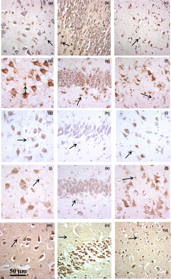
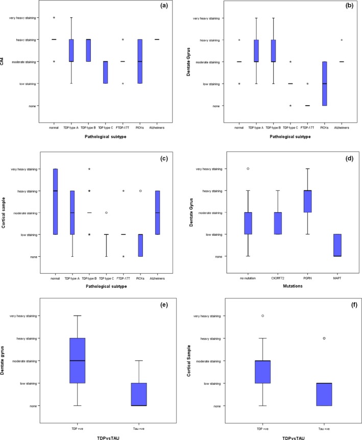
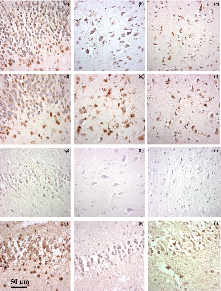
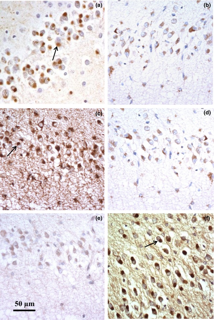
We investigated changes in the degradative pathways in 60 patients with different pathological or genetic forms of FTLD employing immunohistochemistry for marker proteins such as lysosomal-associated membrane proteins 1 (LAMP-1) and 2 (LAMP-2), cathepsin D (CTSD) and microtubule-associated protein 1 light chain 3 alpha (LC3A). Immunostained sections were qualitatively and semi-quantitatively assessed for the appearance, distribution and intensity of staining in neurones of the dentate gyrus (DG) and CA4 region of the hippocampus, and the temporal cortex (Tcx).
Lower levels of neuronal LAMP-1 immunostaining were present in the DG and Tcx in FTLD-tau compared to FTLD-TDP. There was less LAMP-1 immunostaining in FTLD-tau with MAPT mutations, and FTLD-tau with Pick bodies, compared to FTLD-TDP types A and B, and less LAMP-1 immunostaining in FTLD-TDP type C than in FTLD-TDP types A and B. There was greater LAMP-1 immunostaining in GRN mutation which may reflect the underlying type A histology rather than mutation. There were no differences in neuronal LAMP-2, CTSD, EEA-1 or LC3A immunostaining between any of the five FTLD histological or four genetic groups, nor between FTLD-TDP and FTLD-tau.
The underlying pathological mechanism in FTLD-tau may lie with a relative deficiency of lysosomes, or defective vesicular transport, whereas the failure to clear TDP-43 aggregates may lie with lysosomal dysfunction rather than a lack of available lysosomes or degradative enzymes.
细胞生物学和遗传学证据表明,在额颞叶变性(FTLD)的发病机制中,通过自噬或溶酶体途径降解聚集蛋白(如 tau 和 TDP-43)的过程中存在失败。
我们通过免疫组织化学方法研究了 60 例不同病理或遗传形式的 FTLD 患者降解途径的变化,使用溶酶体相关膜蛋白 1(LAMP-1)和 2(LAMP-2)、组织蛋白酶 D(CTSD)和微管相关蛋白 1 轻链 3α(LC3A)等标记蛋白。对齿状回(DG)和海马 CA4 区以及颞叶皮质(Tcx)神经元的染色出现、分布和强度进行了定性和半定量评估。
与 FTLD-TDP 相比,FTLD-tau 中 DG 和 Tcx 神经元的 LAMP-1 免疫染色水平较低。与 FTLD-TDP 类型 A 和 B 相比,MAPT 突变和 Pick 体的 FTLD-tau 中 LAMP-1 免疫染色较少,与 FTLD-TDP 类型 A 和 B 相比,FTLD-TDP 类型 C 中 LAMP-1 免疫染色较少。GRN 突变的 LAMP-1 免疫染色较多,这可能反映了潜在的 A 型组织学,而不是突变。在任何 5 种 FTLD 组织学或 4 种遗传组之间,以及在 FTLD-TDP 和 FTLD-tau 之间,神经元 LAMP-2、CTSD、EEA-1 或 LC3A 免疫染色均无差异。
FTLD-tau 的潜在病理机制可能在于溶酶体相对缺乏,或囊泡运输缺陷,而 TDP-43 聚集物无法清除可能与溶酶体功能障碍有关,而不是缺乏可用的溶酶体或降解酶。